Lewis M J, Olby N J, Early P J, Mariani C L, Muñana K R, Seiler G S, Griffith E H
College of Veterinary Medicine, NCSU, Raleigh, NC.
Comparative Medicine Institute, NCSU, Raleigh, NC.
J Vet Intern Med. 2016 Sep;30(5):1672-1680. doi: 10.1111/jvim.14526. Epub 2016 Sep 12.
Quantification of brain herniation on MRI and its immediate clinical implications are poorly described.
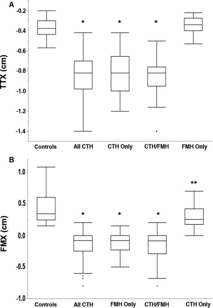
Define the normal position of caudal fossa structures on brain MRIs in dogs and cats utilizing morphometry, compare this to dogs and cats with caudal transtentorial herniation (CTH), foramen magnum herniation (FMH) or both identified on MRI, and investigate associations between herniation severity, clinical signs, and 24-hour outcome.
Ninety-two controls (66 dogs, 26 cats), 119 cases with herniation (88 dogs, 31 cats).
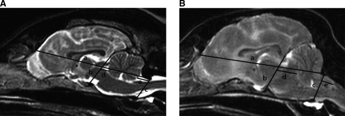
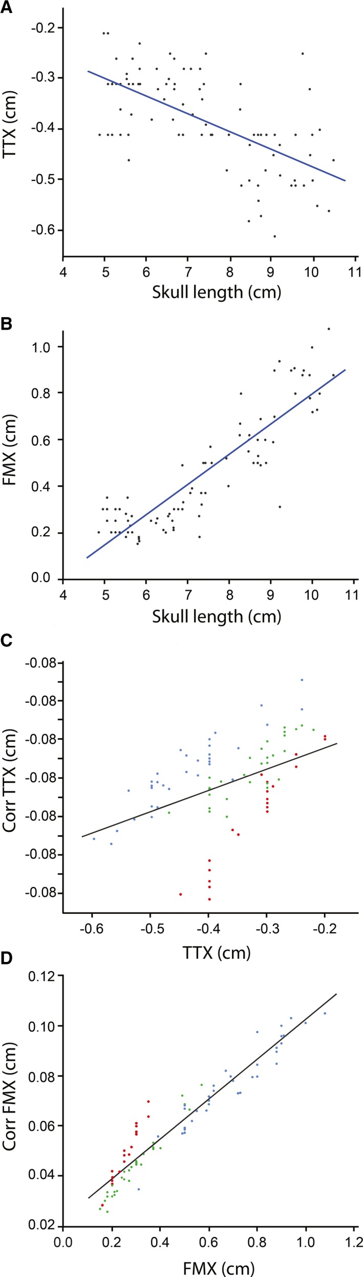
Retrospective case series. The MRI database was searched for controls with normal brain anatomy and cases with brain herniation. Morphometry in controls established TTX (transtentorial to rostroventral cerebellum) to quantify CTH and FMX (caudoventral cerebellum to foramen magnum) to quantify FMH. Measurements were compared between cases and controls. Correlations with specific clinical variables and outcome were investigated.
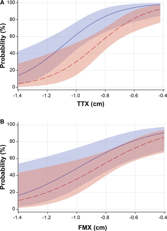
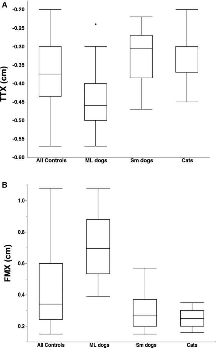
Measurements in medium/large control dogs versus small dog and cat controls were significantly different (P < .001, TTX: -0.46, -0.305, -0.3, FMX: 0.695, 0.27, 0.25, respectively). 119/1564 (7.6%) cases that underwent brain imaging had brain herniation. TTX and FMX were significantly different between controls and cases with CTH or FMH (P < .001). 67/89 (75%) cases with supratentorial lesions had no signs directly attributable to herniation. 71/119 (60%) had a normal anesthetic recovery. TTX was significantly associated with 24-hour survival (P < .001).
Brain herniation can be quantified on MRI. Clinical signs directly attributable to brain herniation commonly are absent, and more severe CTH based on TTX is associated with a worse short-term outcome.
MRI上脑疝的量化及其直接临床意义描述甚少。
利用形态测量法确定犬猫脑部MRI上颅后窝结构的正常位置,将其与MRI上发现有幕下经小脑幕疝(CTH)、枕骨大孔疝(FMH)或两者皆有的犬猫进行比较,并研究疝严重程度、临床症状与24小时预后之间的关联。
92只对照动物(66只犬,26只猫),119例有脑疝的病例(88只犬,31只猫)。
回顾性病例系列研究。在MRI数据库中搜索脑部解剖结构正常的对照动物和有脑疝的病例。对照动物的形态测量确定了TTX(幕下至小脑嘴腹侧)以量化CTH,以及FMX(小脑尾腹侧至枕骨大孔)以量化FMH。对病例和对照动物的测量结果进行比较。研究与特定临床变量和预后的相关性。
中型/大型对照犬与小型犬及猫对照的测量结果有显著差异(P <.001,TTX分别为-0.46、-0.305、-0.3,FMX分别为0.695、0.27、0.25)。1564例接受脑部成像的病例中有119例(7.6%)有脑疝。CTH或FMH病例与对照动物之间的TTX和FMX有显著差异(P <.001)。89例幕上病变病例中有67例(75%)没有直接归因于脑疝的症状。119例中有71例(60%)麻醉恢复正常。TTX与24小时生存率显著相关(P <.001)。
MRI可对脑疝进行量化。通常不存在直接归因于脑疝的临床症状,基于TTX的更严重CTH与更差的短期预后相关。