• 文献检索
  • 文档翻译
  • 深度研究
  • 学术资讯
  • Suppr Zotero 插件Zotero 插件
  • 邀请有礼
  • 套餐&价格
  • 历史记录
应用&插件
Suppr Zotero 插件Zotero 插件浏览器插件Mac 客户端Windows 客户端微信小程序
定价
高级版会员购买积分包购买API积分包
服务
文献检索文档翻译深度研究API 文档MCP 服务
关于我们
关于 Suppr公司介绍联系我们用户协议隐私条款
关注我们

Suppr 超能文献

核心技术专利:CN118964589B侵权必究
粤ICP备2023148730 号-1Suppr @ 2026

文献检索

告别复杂PubMed语法,用中文像聊天一样搜索,搜遍4000万医学文献。AI智能推荐,让科研检索更轻松。

立即免费搜索

文件翻译

保留排版,准确专业,支持PDF/Word/PPT等文件格式,支持 12+语言互译。

免费翻译文档

深度研究

AI帮你快速写综述,25分钟生成高质量综述,智能提取关键信息,辅助科研写作。

立即免费体验

玻璃体内注射贝伐单抗对未经治疗的视网膜分支静脉阻塞所致黄斑水肿中未受影响视网膜的潜在缺血效应分析:一项前瞻性干预性病例系列研究

Analysis of Potential Ischemic Effect of Intravitreal Bevacizumab on Unaffected Retina in Treatment-Naïve Macular Edema Due to Branch Retinal Vein Occlusion: A Prospective, Interventional Case-Series.

作者信息

Rishi Pukhraj, Raka Neha, Rishi Ekta

机构信息

Shri Bhagwan Mahavir Vitreoretinal Services, Sankara Nethralaya, Chennai, TamilNadu, India.

出版信息

PLoS One. 2016 Sep 12;11(9):e0162533. doi: 10.1371/journal.pone.0162533. eCollection 2016.

DOI:10.1371/journal.pone.0162533
PMID:27618696
原文链接:https://pmc.ncbi.nlm.nih.gov/articles/PMC5019367/
Abstract

BACKGROUND

To study potential ischemic effects of intravitreal Bevacizumab (IVB) on unaffected retina in treatment-naive eyes with macular edema secondary to branch retinal vein occlusion (BRVO) and contralateral eyes secondary to systemic absorption.

METHODS AND FINDINGS

Prospective, interventional series included 27 treatment-naive eyes with BRVO and macular edema.

EXCLUSION CRITERIA

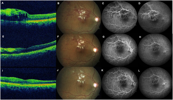
Eyes with diabetic retinopathy, glaucoma, vasculitides, papilledema or systemic neurologic condition. Subjects underwent complete ophthalmological examination including fluoroscein angiography (FA), optical coherence tomography (OCT) and multifocal electroretinogram (mf-ERG). All subjects received single 1.25 mg/0.05ml IVB injection. Two observers measured all parameters; inter-observer agreements were expressed as kappa values. Paired t-test was used to compare values at baseline and follow-up. The statistical analysis was done using SPSS for Windows, Version 14.0. (Chicago, SPSS Inc.) Presenting mean CFT (central foveal thickness) was 499.5(+/-229.7) μm, mean BCVA (best corrected visual acuity) was 0.64(+/-0.41) logMAR. At last follow-up, mean CFT was 267.9(+/-159.3) μm (P<0.001), 95% CI [127.18, 422.32]; mean BCVA was 0.28(+/-0.24) logMAR. Respectively, mean N1 and P1 amplitudes of mfERG in 'unaffected quadrant' at presentation were -6.10(+/-4.00) nV/deg2 and 17.17(+/-11.54)nV/deg2; and -5.33(+/-1.30)nV/deg2 and 15.29(+/-4.69)nV/deg2 at final follow-up (P = 0.631 and 0.197, respectively), (95% CIs [-0.93, 1.42] and [-4.22, 1.08] respectively). In fundus quadrant of fellow eyes corresponding to unaffected quadrant in treated eyes, mean N1 and P1 amplitudes at presentation were -5.39(+/-1.56)nV/deg2 and 15.89(+/-3.89)nV/deg2; and -5.39(+/-1.90)nV/deg2 and 15.9(+/-5.52)nV/deg2 (P = 0.380 and 0.208), (95% CIs [-0.57, 1.28] and [-4.1, 1.1]) at last follow-up, respectively.

LIMITATIONS

This study analysed the effects with a single injection of bevacizumab. However, whether ischemic adverse effects will emerge with repeated IVB injections as a consequence of cumulative dosing needs further investigation. The setting of our study being a tertiary care centre, the numbers of fresh BRVO cases without prior intervention were limited. Thus, the limitations of our study include a small sample size with a small follow-up period. No major ocular/systemic adverse event was observed in the study period.

CONCLUSION

No evidence of progressive ischaemia attributable to single bevacizumab treatment was observed in this study. However, a larger prospective study involving subjects with cumulative dosing of bevacizumab and a longer follow-up could provide a better understanding of the potential ischaemic effects of bevacizumab or other anti-VEGF agents.

https://cdn.ncbi.nlm.nih.gov/pmc/blobs/ac94/5019367/f30ca9b0206b/pone.0162533.g002.jpg
https://cdn.ncbi.nlm.nih.gov/pmc/blobs/ac94/5019367/0de49002a14b/pone.0162533.g001.jpg
https://cdn.ncbi.nlm.nih.gov/pmc/blobs/ac94/5019367/f30ca9b0206b/pone.0162533.g002.jpg
https://cdn.ncbi.nlm.nih.gov/pmc/blobs/ac94/5019367/0de49002a14b/pone.0162533.g001.jpg
https://cdn.ncbi.nlm.nih.gov/pmc/blobs/ac94/5019367/f30ca9b0206b/pone.0162533.g002.jpg
摘要

背景

研究玻璃体内注射贝伐单抗(IVB)对初治的视网膜分支静脉阻塞(BRVO)继发黄斑水肿患眼及对侧眼未受影响视网膜的潜在缺血性影响,以及全身吸收的情况。

方法与结果

前瞻性干预系列研究纳入了27例初治的BRVO继发黄斑水肿患眼。

排除标准

患有糖尿病性视网膜病变、青光眼、血管炎、视乳头水肿或全身性神经系统疾病的眼。受试者接受了包括荧光素血管造影(FA)、光学相干断层扫描(OCT)和多焦视网膜电图(mf-ERG)在内的全面眼科检查。所有受试者均接受了单次1.25mg/0.05ml的IVB注射。两名观察者测量所有参数;观察者间一致性用kappa值表示。配对t检验用于比较基线和随访时的值。使用SPSS for Windows 14.0版(芝加哥,SPSS公司)进行统计分析。呈现的平均中心凹厚度(CFT)为499.5(±229.7)μm,平均最佳矫正视力(BCVA)为0.64(±0.41)logMAR。在最后一次随访时,平均CFT为267.9(±159.3)μm(P<0.001),95%置信区间[127.18, 422.32];平均BCVA为0.28(±0.24)logMAR。在初诊时,“未受影响象限”的mfERG平均N1和P1振幅分别为-6.10(±4.00)nV/deg2和17.17(±11.54)nV/deg2;在最后一次随访时分别为-5.33(±1.30)nV/deg2和15.29(±4.69)nV/deg2(P分别为0.631和0.197),(95%置信区间分别为[-0.93, 1.42]和[-4.22, 1.08])。在治疗眼未受影响象限对应的对侧眼眼底象限,初诊时平均N1和P1振幅分别为-5.39(±1.56)nV/deg2和15.89(±3.89)nV/deg2;在最后一次随访时分别为-5.39(±1.90)nV/deg2和15.9(±5.52)nV/deg2(P分别为0.380和0.208),(95%置信区间分别为[-0.57, 1.28]和[-4.1, 1.1])。

局限性

本研究分析了单次注射贝伐单抗的效果。然而,由于累积给药,重复IVB注射是否会出现缺血性不良反应需要进一步研究。我们的研究是在三级医疗中心进行的,未经事先干预 的新鲜BRVO病例数量有限。因此,我们研究的局限性包括样本量小和随访期短。在研究期间未观察到重大眼/全身不良事件。

结论

本研究未观察到单次贝伐单抗治疗导致进行性缺血的证据。然而,一项涉及贝伐单抗累积给药受试者且随访期更长的更大规模前瞻性研究,可能会更好地了解贝伐单抗或其他抗VEGF药物的潜在缺血性影响。

相似文献

1
Analysis of Potential Ischemic Effect of Intravitreal Bevacizumab on Unaffected Retina in Treatment-Naïve Macular Edema Due to Branch Retinal Vein Occlusion: A Prospective, Interventional Case-Series.玻璃体内注射贝伐单抗对未经治疗的视网膜分支静脉阻塞所致黄斑水肿中未受影响视网膜的潜在缺血效应分析:一项前瞻性干预性病例系列研究
PLoS One. 2016 Sep 12;11(9):e0162533. doi: 10.1371/journal.pone.0162533. eCollection 2016.
2
Clinical, anatomical, and electrophysiological assessments of the central retina following intravitreal bevacizumab for macular edema secondary to retinal vein occlusion.玻璃体内注射贝伐单抗治疗视网膜静脉阻塞继发黄斑水肿后对视网膜中央的临床、解剖学及电生理评估
Int Ophthalmol. 2016 Feb;36(1):21-36. doi: 10.1007/s10792-015-0066-6. Epub 2015 Mar 29.
3
"Off-label" use of intravitreal bevacizumab in non-ischemic macular edema secondary to retinal vein obstructions.玻璃体内注射贝伐单抗在视网膜静脉阻塞继发的非缺血性黄斑水肿中的“超说明书”使用。
Rom J Ophthalmol. 2016 Apr-Jun;60(2):90-95.
4
Prospective study of intravitreal triamcinolone acetonide versus bevacizumab for macular edema secondary to central retinal vein occlusion.前瞻性研究曲安奈德与贝伐单抗治疗视网膜中央静脉阻塞继发黄斑水肿。
Retina. 2011 May;31(5):838-45. doi: 10.1097/IAE.0b013e3181f4420d.
5
Combination therapy with intravitreal bevacizumab and macular grid and scatter laser photocoagulation in patients with macular edema secondary to branch retinal vein occlusion.玻璃体腔注射贝伐单抗联合黄斑格栅与多点散射激光光凝治疗视网膜分支静脉阻塞继发黄斑水肿患者
J Ocul Pharmacol Ther. 2015 Apr;31(3):179-85. doi: 10.1089/jop.2014.0069. Epub 2015 Feb 25.
6
Predictive factors for functional improvement after intravitreal bevacizumab therapy for macular edema due to branch retinal vein occlusion.预测因子分析:玻璃体内注射贝伐单抗治疗视网膜分支静脉阻塞所致黄斑水肿的功能改善。
Graefes Arch Clin Exp Ophthalmol. 2011 Feb;249(2):183-92. doi: 10.1007/s00417-010-1470-2. Epub 2010 Aug 18.
7
Clinical, anatomic, and electrophysiologic evaluation following intravitreal bevacizumab for macular edema in retinal vein occlusion.玻璃体内注射贝伐单抗治疗视网膜静脉阻塞所致黄斑水肿后的临床、解剖学及电生理学评估
Am J Ophthalmol. 2007 Apr;143(4):601-6. doi: 10.1016/j.ajo.2006.12.037. Epub 2007 Feb 15.
8
Intravitreal ranibizumab (Lucentis) for branch retinal vein occlusion-induced macular edema: nine-month results of a prospective study.玻璃体内雷珠单抗(Lucentis)治疗分支静脉阻塞相关性黄斑水肿:一项前瞻性研究的九个月结果。
Retina. 2010 Jun;30(6):893-902. doi: 10.1097/IAE.0b013e3181cd4894.
9
EFFICACY AND FREQUENCY OF INTRAVITREAL AFLIBERCEPT VERSUS BEVACIZUMAB FOR MACULAR EDEMA SECONDARY TO CENTRAL RETINAL VEIN OCCLUSION.阿柏西普与雷珠单抗治疗视网膜中央静脉阻塞继发黄斑水肿的疗效及频率。
Retina. 2018 Sep;38(9):1795-1800. doi: 10.1097/IAE.0000000000001782.
10
Extrafoveal changes following intravitreal bevacizumab injections for macular edema secondary to branch retinal vein occlusion: an mfERG and OCT study.玻璃体腔注射贝伐单抗治疗视网膜分支静脉阻塞继发黄斑水肿后的黄斑外改变:一项多焦视网膜电图和光学相干断层扫描研究
Doc Ophthalmol. 2013 Apr;126(2):137-48. doi: 10.1007/s10633-012-9367-5. Epub 2012 Dec 20.

引用本文的文献

1
Five-year outcomes of retinal vein occlusion treated with vascular endothelial growth factor inhibitors.血管内皮生长因子抑制剂治疗视网膜静脉阻塞的五年疗效
BMJ Open Ophthalmol. 2019 Mar 29;4(1):e000249. doi: 10.1136/bmjophth-2018-000249. eCollection 2019.
2
Macular function following intravitreal ranibizumab for macular edema associated with branch retinal vein occlusion.玻璃体内注射雷珠单抗治疗视网膜分支静脉阻塞相关黄斑水肿后的黄斑功能
Doc Ophthalmol. 2019 Aug;139(1):67-74. doi: 10.1007/s10633-019-09696-5. Epub 2019 Apr 12.
3
Branch Retinal Vein Occlusion, Macular Ischemia, and Intravitreal Anti-VEGF Therapy.

本文引用的文献

1
In vivo detection of acute ischemic damages in retinal arterial occlusion with optical coherence tomography: a "prominent middle limiting membrane sign".光学相干断层扫描对视网膜动脉阻塞患者急性缺血性损伤的活体检测:一种“显著中层膜限征”。
Retina. 2013 Nov-Dec;33(10):2110-7. doi: 10.1097/IAE.0b013e3182899205.
2
Accelerated ischemic vascular retinopathy after intravitreally injected bevacizumab for central retinal vein occlusion in elderly patients.老年患者玻璃体内注射贝伐单抗治疗视网膜中央静脉阻塞后加速性缺血性视网膜病变
Clin Ophthalmol. 2013;7:455-60. doi: 10.2147/OPTH.S30156. Epub 2013 Mar 1.
3
Adverse events with intravitreal injection of vascular endothelial growth factor inhibitors: nested case-control study.
视网膜分支静脉阻塞、黄斑缺血与玻璃体内抗血管内皮生长因子治疗
Case Rep Ophthalmol. 2017 Apr 28;8(1):271-278. doi: 10.1159/000475520. eCollection 2017 Jan-Apr.
玻璃体内注射血管内皮生长因子抑制剂的不良反应:巢式病例对照研究。
BMJ. 2012 Jul 4;345:e4203. doi: 10.1136/bmj.e4203.
4
Sustained benefits from ranibizumab for macular edema following branch retinal vein occlusion: 12-month outcomes of a phase III study.雷珠单抗治疗视网膜分支静脉阻塞继发黄斑水肿的持续获益:一项 III 期研究的 12 个月结果。
Ophthalmology. 2011 Aug;118(8):1594-602. doi: 10.1016/j.ophtha.2011.02.022.
5
Autofluorescence and high-definition optical coherence tomography of retinal artery occlusions.视网膜动脉阻塞的自体荧光与高清光学相干断层扫描
Clin Ophthalmol. 2010 Oct 21;4:1159-63. doi: 10.2147/OPTH.S13592.
6
Inner neural retina loss in central retinal artery occlusion.中心性视网膜动脉阻塞的内层神经视网膜丧失。
Jpn J Ophthalmol. 2010 Sep;54(5):423-9. doi: 10.1007/s10384-010-0841-x. Epub 2010 Nov 5.
7
Ischemic maculopathy following intravitreal bevacizumab for refractory diabetic macular edema.
Int Ophthalmol. 2011 Feb;31(1):39-42. doi: 10.1007/s10792-010-9390-z. Epub 2010 Sep 18.
8
Efficacy and safety of anti-vascular endothelial growth factor (VEGF) therapy with intravitreal ranibizumab (Lucentis) for naive retinal vein occlusion: 1-year follow-up.抗血管内皮生长因子(VEGF)治疗药物雷珠单抗(Lucentis)治疗初发性视网膜静脉阻塞的疗效和安全性:1 年随访。
Br J Ophthalmol. 2011 Jan;95(1):56-68. doi: 10.1136/bjo.2009.174060. Epub 2010 Jul 3.
9
Ranibizumab for macular edema following branch retinal vein occlusion: six-month primary end point results of a phase III study.雷珠单抗治疗视网膜分支静脉阻塞继发黄斑水肿:一项 III 期研究的 6 个月主要终点结果。
Ophthalmology. 2010 Jun;117(6):1102-1112.e1. doi: 10.1016/j.ophtha.2010.02.021. Epub 2010 Apr 15.
10
Ranibizumab for macular edema following central retinal vein occlusion: six-month primary end point results of a phase III study.雷珠单抗治疗视网膜中央静脉阻塞继发黄斑水肿:一项 III 期研究的 6 个月主要终点结果。
Ophthalmology. 2010 Jun;117(6):1124-1133.e1. doi: 10.1016/j.ophtha.2010.02.022. Epub 2010 Apr 9.