Roy Soumen, Pavitrakar Daya, Gunjikar Rashmi, Ayachit Vijay M, Bondre Vijay P, Sapkal Gajanan N
National Institute of Virology, Sus Road, Pashan, Pune, 411021, India.
BMC Infect Dis. 2016 Sep 14;16:487. doi: 10.1186/s12879-016-1794-6.
Interaction between immune system and Chandipura virus (CHPV) during different stages of its life cycle remain poorly understood. The exact route of virus entry into the blood and CNS invasion has not been clearly defined. The present study was undertaken to assess the population in PBMC that supports the growth of virus and to detect active virus replication in PBMC as well as its subsets.
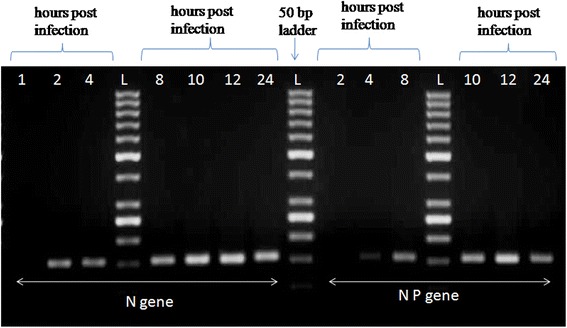
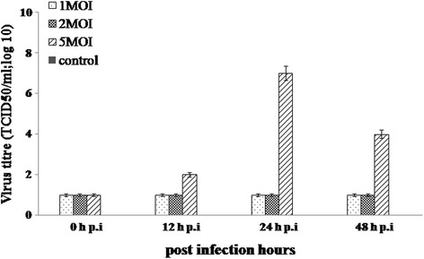
PBMC subsets viz.: CD3(+), CD14(+), CD19(+), CD56(+)cells were separated and infected with CHPV. The infected cells were then assessed for transcription (N gene primer) and replication (NP gene primer) of CHPV by PCR. The supernatant collected from infected cells were titrated in Baby Hamster Kidney (BHK) cells to assess virus release. The cytokine and chemokine expression was quantified by flow cytometry.
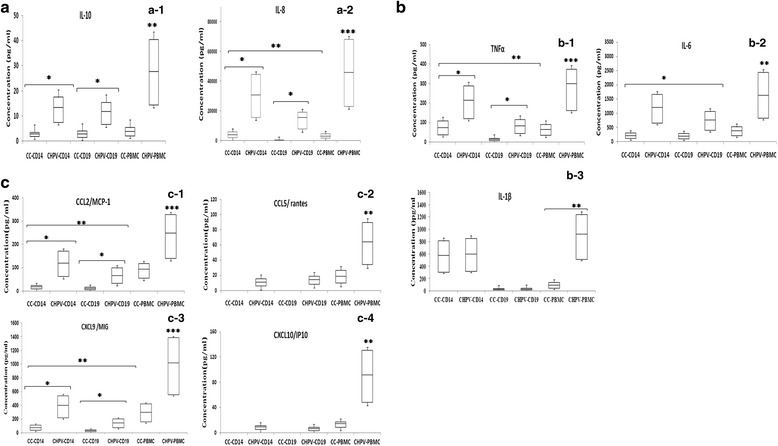
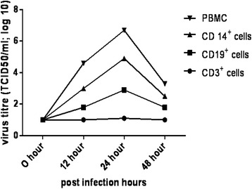
Amplification of N and NP gene was detected in CD14(+) (monocyte) and CD19(+) (B cell), significant increase in virus titre was also observed in these subsets. It was observed that, although the levels of IL-6 and IL-10 were elevated in CD14(+) cells as compared to CD19(+)cells, the differences were not significant. However the levels of TNFα and IL-8 were significantly elevated in CD14(+) cells than in CD19(+)cells. The levels of chemokine (CXCL9, CCL5, CCL2, CXCL10) were significantly elevated in CHPV infected PBMC as compared to uninfected cells. CCL2 and CXCL9 were significantly increased in CHPV infected CD14(+)cells as compared to CD19(+) cells.
CD14(+)and CD19(+)cells support active replication of CHPV. High viral load was detected in CD14(+) cells infected with CHPV hence it might be the primary target cells for active replication of CHPV. An elevated levels of cytokines and chemokines observed in CD14(+) cells may help in predicting the pathogenecity of CHPV and possible entry into the central nervous system.
免疫系统与钱德普拉病毒(CHPV)在其生命周期不同阶段的相互作用仍知之甚少。病毒进入血液的确切途径以及中枢神经系统侵袭尚未明确界定。本研究旨在评估支持病毒生长的外周血单个核细胞(PBMC)群体,并检测PBMC及其亚群中的活跃病毒复制。
分离PBMC亚群,即CD3(+)、CD14(+)、CD19(+)、CD56(+)细胞,并用CHPV感染。然后通过聚合酶链反应(PCR)评估感染细胞中CHPV的转录(N基因引物)和复制(NP基因引物)。从感染细胞收集的上清液在幼仓鼠肾(BHK)细胞中进行滴定以评估病毒释放。通过流式细胞术对细胞因子和趋化因子表达进行定量。
在CD14(+)(单核细胞)和CD19(+)(B细胞)中检测到N和NP基因的扩增,在这些亚群中也观察到病毒滴度显著增加。据观察,虽然与CD19(+)细胞相比,CD14(+)细胞中白细胞介素-6(IL-6)和白细胞介素-10(IL-10)水平升高,但差异不显著。然而,CD14(+)细胞中肿瘤坏死因子α(TNFα)和白细胞介素-8(IL-8)水平比CD19(+)细胞中显著升高。与未感染细胞相比,CHPV感染的PBMC中趋化因子(CXCL9、CCL5、CCL2、CXCL10)水平显著升高。与CD19(+)细胞相比,CHPV感染的CD14(+)细胞中CCL2和CXCL9显著增加。
CD14(+)和CD19(+)细胞支持CHPV的活跃复制。在感染CHPV的CD14(+)细胞中检测到高病毒载量,因此它可能是CHPV活跃复制的主要靶细胞。在CD14(+)细胞中观察到的细胞因子和趋化因子水平升高可能有助于预测CHPV的致病性以及进入中枢神经系统的可能性。