Chandipura Virus Group, National Institute of Virology, Pune-411001, India.
Virol J. 2011 May 25;8:259. doi: 10.1186/1743-422X-8-259.
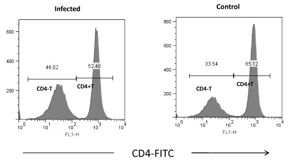
Chandipura virus produces acute infection in mice. During infection drastic reduction of CD4+, CD8+ and CD19 + cell was noticed. Depletion of lymphocytes also noticed in spleen. The reduction may be due to the regulatory mechanism of immune system to prevent the bystander host tissue injury. There are several mechanisms like generation of regulatory cells, activation induced cell death (ACID) etc were indicated to control the activation and maintain cellular homeostasis. Role of regulatory cells in homeostasis has been described in several viral diseases. This study was undertaken to characterize CD4+T regulatory cells from the infected mice.
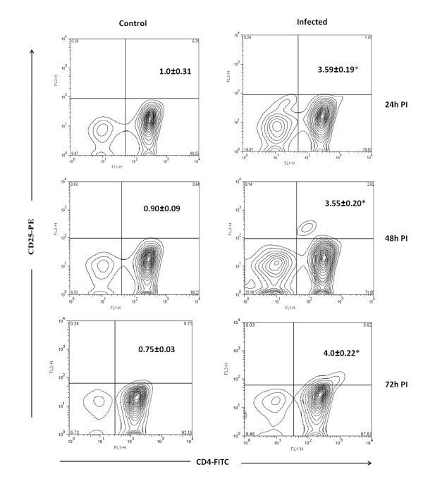
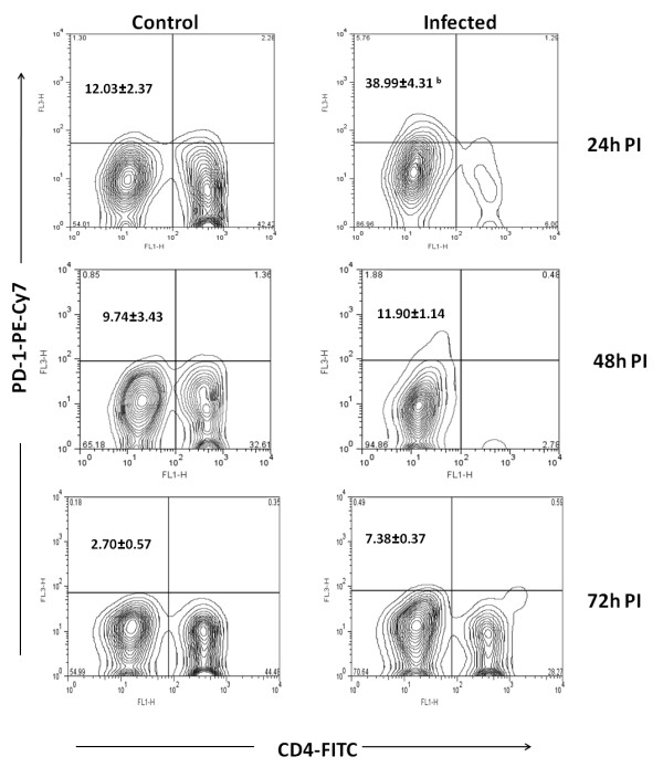
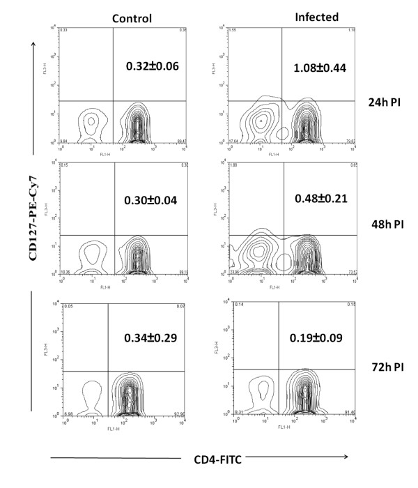
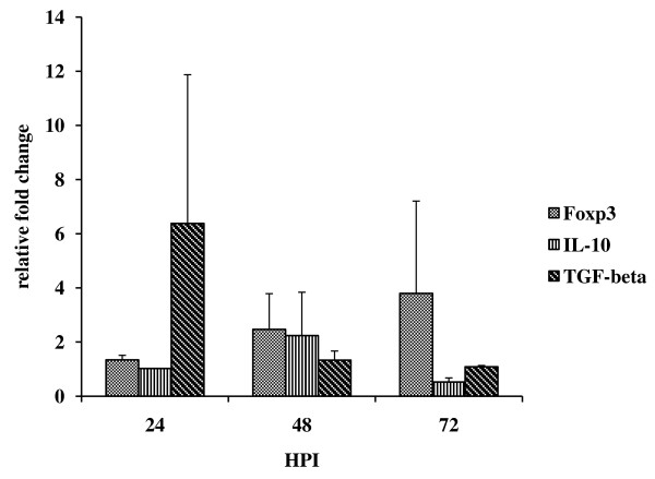
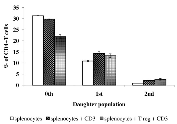
In this study we purified the CD4+ T cells from Chandipura virus infected susceptible Balb/c mice. CD4+ T regulatory cells were identified by expression of cell surface markers CD25, CD127 and CTLA-4 and intracellular markers Foxp3, IL-10 and TGF-beta. Antigen specificity and ability to suppress the proliferation of other lymphocytes were studied in vitro by purified CD4+CD25+T regulatory cells from infected mice. The proliferation was calculated by proliferation module of Flow Jo software. Expression of death receptors on regulatory cells were studied by flowcytometer.
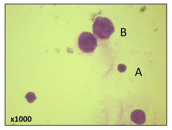
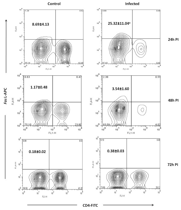
The CD4+ T cells isolated from infected mice expressed characteristic markers of regulatory phenotype at all post infective hours tested. The CD4+ T regulatory cells were proliferated when stimulated with Chandipura virus antigen. The regulatory cells did not suppress the proliferation of splenocytes stimulated with anti CD3 antibody when co cultured with them. Interesting observation was, while purification of CD4+ T cells by negative selection, the population of cells negative for CD4 also co purified along with CD4+ T cell. Flow cytometry analysis and light microscopy revealed that CD4 negative cells were of different size and shape (atypical) compared to the normal lymphocytes. Greater percentage of these atypical lymphocytes expressed Fas Ligand and Programmed Death1 (PD-1) receptor.
From these results we concluded that virus specific CD4+T regulatory cells are generated during Chandipura virus infection in mice and these cells might control the activated lymphocytes during infection by different mechanism.
钱德拉普尔病毒会在老鼠体内引起急性感染。在感染过程中,观察到 CD4+、CD8+ 和 CD19+细胞急剧减少。脾脏中的淋巴细胞也被耗尽。这种减少可能是由于免疫系统的调节机制,以防止旁观者宿主组织损伤。有几种机制,如调节细胞的产生、激活诱导的细胞死亡(ACID)等,被认为可以控制激活并维持细胞内稳态。调节细胞在体内平衡中的作用已在几种病毒性疾病中得到描述。本研究旨在从感染的老鼠中鉴定 Chandipura 病毒感染的调节性 CD4+T 细胞。
在这项研究中,我们从 Chandipura 病毒易感的 Balb/c 感染的老鼠中纯化了 CD4+T 细胞。通过表达细胞表面标志物 CD25、CD127 和 CTLA-4 以及细胞内标志物 Foxp3、IL-10 和 TGF-β,鉴定了 CD4+T 调节细胞。通过从感染的老鼠中纯化 CD4+CD25+T 调节细胞,在体外研究了抗原特异性和抑制其他淋巴细胞增殖的能力。增殖通过 Flow Jo 软件的增殖模块进行计算。通过流式细胞仪研究了调节细胞上死亡受体的表达。
从感染的老鼠中分离的 CD4+T 细胞在所有检测的感染后时间点均表达调节表型的特征性标志物。当用 Chandipura 病毒抗原刺激时,CD4+T 调节细胞增殖。当与抗 CD3 抗体刺激的脾细胞共培养时,调节细胞不会抑制其增殖。有趣的观察是,在用阴性选择纯化 CD4+T 细胞时,CD4 阴性细胞也与 CD4+T 细胞一起共同纯化。流式细胞术分析和相差显微镜显示,与正常淋巴细胞相比,CD4 阴性细胞的大小和形状不同(非典型)。这些非典型淋巴细胞中更大比例表达 Fas 配体和程序性死亡 1(PD-1)受体。
从这些结果中我们得出结论,在 Chandipura 病毒感染的老鼠中产生了病毒特异性 CD4+T 调节细胞,这些细胞可能通过不同的机制在感染过程中控制激活的淋巴细胞。