Trauma and Orthopaedics Department, 5th Floor St James' Wing, St George's University Hospitals NHS Foundation Trust, Blackshaw Road, London, SW17 0QT, UK.
St George's, University of London, London, SW17 0RE, UK.
Knee Surg Sports Traumatol Arthrosc. 2017 Dec;25(12):3755-3772. doi: 10.1007/s00167-016-4272-1. Epub 2016 Sep 8.
The medial patellofemoral ligament (MPFL) is the major medial soft-tissue stabiliser of the patella, originating from the medial femoral condyle and inserting onto the medial patella. The exact position reported in the literature varies. Understanding the true anatomical origin and insertion of the MPFL is critical to successful reconstruction. The purpose of this systematic review was to determine these locations.
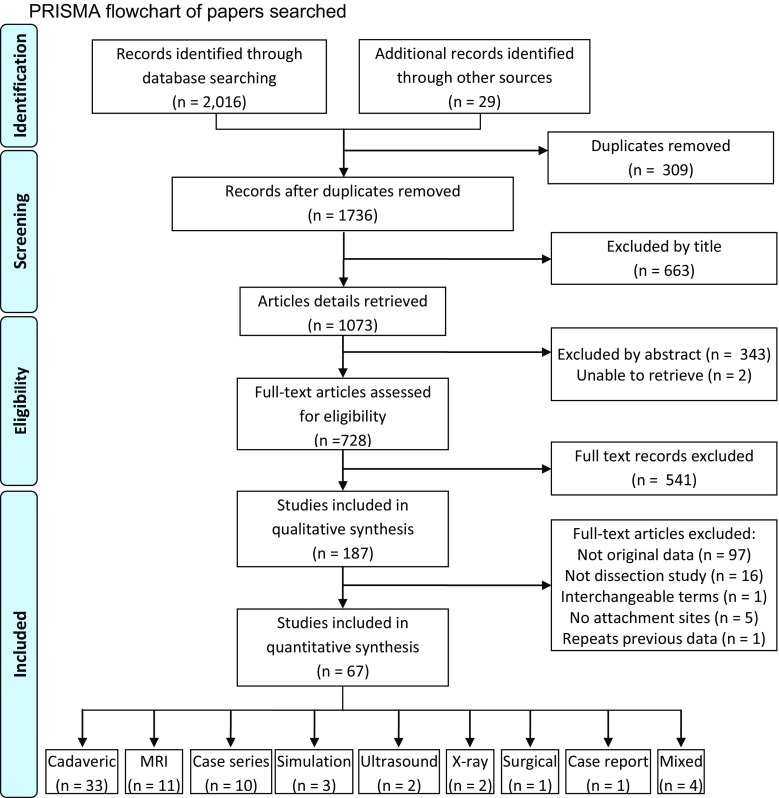
A systematic search of published (AMED, CINAHL, MEDLINE, EMBASE, PubMed and Cochrane Library) and unpublished literature databases was conducted from their inception to the 3 February 2016. All papers investigating the anatomy of the MPFL were eligible. Methodological quality was assessed using a modified CASP tool. A narrative analysis approach was adopted to synthesise the findings.
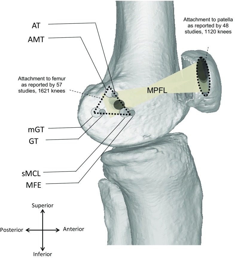
After screening and review of 2045 papers, a total of 67 studies investigating the relevant anatomy were included. From this, the origin appears to be from an area rather than (as previously reported) a single point on the medial femoral condyle. The weighted average length was 56 mm with an 'hourglass' shape, fanning out at both ligament ends.
The MPFL is an hourglass-shaped structure running from a triangular space between the adductor tubercle, medial femoral epicondyle and gastrocnemius tubercle and inserts onto the superomedial aspect of the patella. Awareness of anatomy is critical for assessment, anatomical repair and successful surgical patellar stabilisation.
Systematic review of anatomical dissections and imaging studies, Level IV.
内侧髌股韧带(MPFL)是髌股关节的主要内侧软组织稳定结构,起自股骨内髁,止于髌骨内上缘。文献报道的其确切位置有所不同。了解 MPFL 的真正解剖起点和止点对于成功重建至关重要。本系统评价的目的是确定这些位置。
从建库至 2016 年 2 月 3 日,对已发表(AMED、CINAHL、MEDLINE、EMBASE、PubMed 和 Cochrane Library)和未发表文献数据库进行了系统检索。所有调查 MPFL 解剖结构的论文均符合纳入标准。使用改良的 CASP 工具评估方法学质量。采用叙述性分析方法综合研究结果。
经过筛选和 2045 篇论文的回顾,共有 67 项研究调查了相关解剖结构。由此可见,其起点似乎是在股骨内髁的一个区域,而不是(如先前报道的)一个单点。加权平均长度为 56mm,呈沙漏形,在韧带两端展开。
MPFL 是一种从收肌结节、股骨内上髁和腓肠肌结节之间的三角形空间延伸到髌骨上内侧面的沙漏状结构。了解解剖结构对于评估、解剖修复和成功的手术髌骨稳定至关重要。
解剖学研究的系统评价和影像学研究,IV 级。