Greulich Simon, Arai Andrew E, Sechtem Udo, Mahrholdt Heiko
Division of Cardiology, Robert Bosch Medical Center, Stuttgart, Germany.
National Heart, Lung, and Blood Institute, National Institutes of Health, Department of Health and Human Services, Bethesda, MD, USA.
F1000Res. 2016 Sep 7;5. doi: 10.12688/f1000research.8383.1. eCollection 2016.
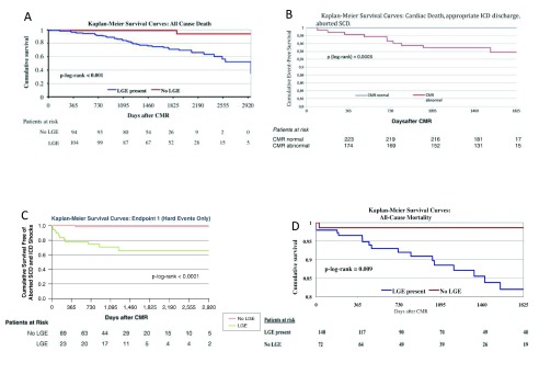
Cardiac magnetic resonance (CMR) is a non-invasive imaging modality that has rapidly emerged during the last few years and has become a valuable, well-established clinical tool. Beside the evaluation of anatomy and function, CMR has its strengths in providing detailed non-invasive myocardial tissue characterization, for which it is considered the current diagnostic gold standard. Late gadolinium enhancement (LGE), with its capability to detect necrosis and to separate ischemic from non-ischemic cardiomyopathies by distinct LGE patterns, offers unique clinical possibilities. The presence of LGE has also proven to be a good predictor of an adverse outcome in various studies. T2-weighted (T2w) images, which are supposed to identify areas of edema and inflammation, are another CMR approach to tissue characterization. However, T2w images have not held their promise owing to several technical limitations and potential physiological concerns. Newer mapping techniques may overcome some of these limitations: they assess quantitatively myocardial tissue properties in absolute terms and show promising results in studies for characterization of diffuse fibrosis (T1 mapping) and/or inflammatory processes (T2 mapping). However, these techniques are still research tools and are not part of the clinical routine yet. T2* CMR has had significant impact in the management of thalassemia because it is possible to image the amount of iron in the heart and the liver, improving both diagnostic imaging and the management of patients with thalassemia. CMR findings frequently have clinical impact on further patient management, and CMR seems to be cost effective in the clinical routine.
心脏磁共振成像(CMR)是一种非侵入性成像方式,在过去几年中迅速兴起,已成为一种有价值的、成熟的临床工具。除了评估解剖结构和功能外,CMR在提供详细的非侵入性心肌组织特征方面具有优势,为此它被认为是当前的诊断金标准。延迟钆增强(LGE)能够检测坏死,并通过不同的LGE模式区分缺血性和非缺血性心肌病,提供了独特的临床可能性。在各种研究中,LGE的存在也已被证明是不良预后的良好预测指标。T2加权(T2w)图像旨在识别水肿和炎症区域,是CMR进行组织特征分析的另一种方法。然而,由于一些技术限制和潜在的生理问题,T2w图像并未达到预期效果。更新的成像技术可能会克服其中一些限制:它们以绝对值定量评估心肌组织特性,并且在弥漫性纤维化(T1成像)和/或炎症过程(T2成像)特征分析的研究中显示出有前景的结果。然而,这些技术仍然是研究工具,尚未成为临床常规检查的一部分。T2* CMR在地中海贫血的管理中产生了重大影响,因为可以对心脏和肝脏中的铁含量进行成像,改善了地中海贫血患者的诊断成像和管理。CMR检查结果常常对患者的进一步管理产生临床影响,并且CMR在临床常规检查中似乎具有成本效益。