• 文献检索
  • 文档翻译
  • 深度研究
  • 学术资讯
  • Suppr Zotero 插件Zotero 插件
  • 邀请有礼
  • 套餐&价格
  • 历史记录
应用&插件
Suppr Zotero 插件Zotero 插件浏览器插件Mac 客户端Windows 客户端微信小程序
定价
高级版会员购买积分包购买API积分包
服务
文献检索文档翻译深度研究API 文档MCP 服务
关于我们
关于 Suppr公司介绍联系我们用户协议隐私条款
关注我们

Suppr 超能文献

核心技术专利:CN118964589B侵权必究
粤ICP备2023148730 号-1Suppr @ 2026

文献检索

告别复杂PubMed语法,用中文像聊天一样搜索,搜遍4000万医学文献。AI智能推荐,让科研检索更轻松。

立即免费搜索

文件翻译

保留排版,准确专业,支持PDF/Word/PPT等文件格式,支持 12+语言互译。

免费翻译文档

深度研究

AI帮你快速写综述,25分钟生成高质量综述,智能提取关键信息,辅助科研写作。

立即免费体验

婴儿主动脉缩窄的磁共振成像:高场强的应用

Magnetic resonance imaging of coarctation of the aorta in infants: use of a high field strength.

作者信息

Baker E J, Ayton V, Smith M A, Parsons J M, Maisey M N, Ladusans E J, Anderson R H, Tynan M, Yates A K, Deverall P B

机构信息

Department of Paediatric Cardiology, Guy's Hospital, London.

出版信息

Br Heart J. 1989 Aug;62(2):97-101. doi: 10.1136/hrt.62.2.97.

DOI:10.1136/hrt.62.2.97
PMID:2765332
原文链接:https://pmc.ncbi.nlm.nih.gov/articles/PMC1216741/
Abstract

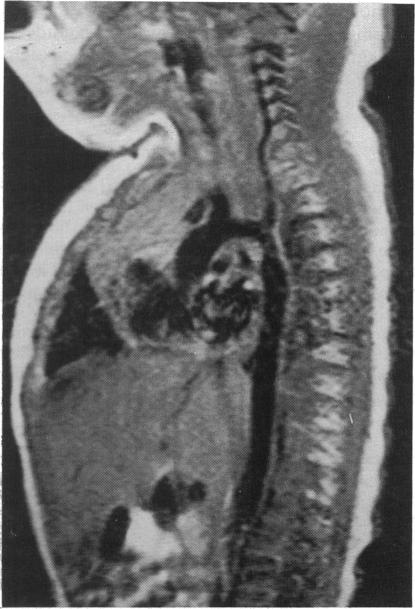
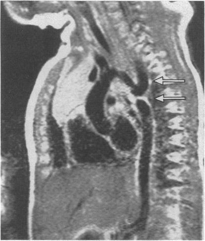
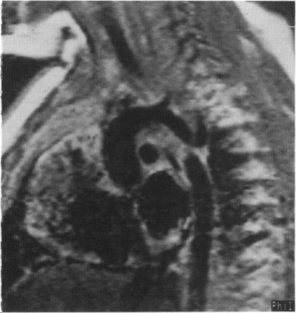
Nineteen infants with suspected coarctation of the aorta were studied with electrocardiographically gated magnetic resonance imaging on a 1.5 T whole body imaging system. In all cases imaging was successful and produced diagnostic images of high resolution. Coarctation was shown in 12 cases. The position and shape of the coarctation were well displayed by the magnetic resonance images. In addition, they clearly showed the relation of the coarctation to arteries arising from the aortic arch and to the length and diameter of the aortic isthmus and the distal aortic arch. The anatomy was confirmed at operation in all 12 patients, except for two with a small ductus arteriosus (arterial duct), which was not seen in the magnetic resonance images. In the seven remaining patients, coarctation was excluded. Magnetic resonance imaging produced high quality images that showed the anatomy better than other non-invasive methods. It provided all the anatomical information required for surgical correction.

摘要

对19例疑似主动脉缩窄的婴儿,在1.5T全身成像系统上采用心电图门控磁共振成像进行研究。所有病例成像均成功,并生成了高分辨率的诊断图像。12例显示有主动脉缩窄。磁共振图像很好地显示了缩窄的位置和形态。此外,它们还清晰地显示了缩窄与主动脉弓发出的动脉、主动脉峡部及主动脉弓远端的长度和直径之间的关系。除2例有小动脉导管(动脉导管)在磁共振图像中未显示外,所有12例患者的解剖结构均在手术中得到证实。其余7例患者排除了主动脉缩窄。磁共振成像生成的高质量图像比其他非侵入性方法能更好地显示解剖结构。它提供了手术矫正所需的所有解剖信息。

https://cdn.ncbi.nlm.nih.gov/pmc/blobs/ec37/1216741/e9bbf6de309f/brheartj00068-0020-a.jpg
https://cdn.ncbi.nlm.nih.gov/pmc/blobs/ec37/1216741/f48fc8b772ad/brheartj00068-0018-a.jpg
https://cdn.ncbi.nlm.nih.gov/pmc/blobs/ec37/1216741/7814267525fe/brheartj00068-0019-a.jpg
https://cdn.ncbi.nlm.nih.gov/pmc/blobs/ec37/1216741/bb145f48d226/brheartj00068-0019-b.jpg
https://cdn.ncbi.nlm.nih.gov/pmc/blobs/ec37/1216741/f273f32ebaed/brheartj00068-0019-c.jpg
https://cdn.ncbi.nlm.nih.gov/pmc/blobs/ec37/1216741/e9bbf6de309f/brheartj00068-0020-a.jpg

相似文献

1
Magnetic resonance imaging of coarctation of the aorta in infants: use of a high field strength.婴儿主动脉缩窄的磁共振成像:高场强的应用
Br Heart J. 1989 Aug;62(2):97-101. doi: 10.1136/hrt.62.2.97.
2
Coarctation and other obstructive aortic arch anomalies: their relationship to the ductus arteriosus.主动脉缩窄及其他主动脉弓梗阻性畸形:它们与动脉导管的关系。
Int J Cardiol. 1986 Dec;13(3):289-308. doi: 10.1016/0167-5273(86)90116-6.
3
Reconstruction of hypoplastic aortic arch and coarctation in a 720-gram premature infant.一名体重720克的早产儿主动脉弓发育不全及缩窄的重建手术
J Pediatr Surg. 1993 May;28(5):731-4. doi: 10.1016/0022-3468(93)90044-l.
4
Coarctation of a right-sided aortic arch and an unusual vascular ring.右侧主动脉弓缩窄及一种不寻常的血管环。
J Cardiovasc Magn Reson. 2004;6(2):577-81. doi: 10.1081/jcmr-120030590.
5
Ascending-descending aortic bypass surgery in aortic arch coarctation: four-dimensional magnetic resonance flow analysis.升主动脉-降主动脉旁路手术治疗主动脉弓缩窄:四维磁共振血流分析
J Thorac Cardiovasc Surg. 2007 Jan;133(1):260-2. doi: 10.1016/j.jtcvs.2006.08.061.
6
Treatment of complex coarctation of aorta with hypoplastic transverse aortic arch using left carotid artery flap.使用左颈动脉瓣治疗合并主动脉弓发育不全的复杂性主动脉缩窄。
Interact Cardiovasc Thorac Surg. 2009 May;8(5):581-3. doi: 10.1510/icvts.2008.197178. Epub 2009 Feb 25.
7
Coarctation, tubular hypoplasia, and the ductus arteriosus. Histological study of 35 specimens.缩窄、管状发育不全与动脉导管。35例标本的组织学研究。
Br Heart J. 1979 Mar;41(3):268-74. doi: 10.1136/hrt.41.3.268.
8
Quantitative morphology of the aortic arch in neonatal coarctation.新生儿主动脉缩窄时主动脉弓的定量形态学
J Am Coll Cardiol. 1986 Sep;8(3):616-20. doi: 10.1016/s0735-1097(86)80191-7.
9
Velocity-encoded magnetic resonance image assessment of regional aortic flow in coarctation patients.缩窄患者局部主动脉血流的速度编码磁共振成像评估
Ann Thorac Surg. 2006 Mar;81(3):1002-7. doi: 10.1016/j.athoracsur.2005.07.002.
10
Coarctation repair using end-to-side anastomosis of descending aorta to proximal aortic arch.采用降主动脉与主动脉弓近端端侧吻合术进行缩窄修复。
Ann Thorac Surg. 1996 Mar;61(3):840-4. doi: 10.1016/0003-4975(95)01153-6.

引用本文的文献

1
Evaluation of aortic coarctation after surgical repair: role of magnetic resonance imaging and Doppler ultrasound.手术修复后主动脉缩窄的评估:磁共振成像和多普勒超声的作用
Br Heart J. 1993 Sep;70(3):285-90. doi: 10.1136/hrt.70.3.285.
2
Magnetic resonance imaging at a high field strength of ventricular septal defects in infants.婴儿室间隔缺损的高场强磁共振成像
Br Heart J. 1989 Oct;62(4):305-10. doi: 10.1136/hrt.62.4.305.
3
Morphological evaluation of atrioventricular septal defects by magnetic resonance imaging.磁共振成像对房室间隔缺损的形态学评估

本文引用的文献

1
Cross-sectional echocardiographic assessment of coarctation in the sick neonate and infant.患病新生儿和婴儿主动脉缩窄的横断面超声心动图评估。
Br Heart J. 1983 Oct;50(4):349-61. doi: 10.1136/hrt.50.4.349.
2
Coarctation of the aorta in infancy.
Circulation. 1969 Sep;40(3):385-98. doi: 10.1161/01.cir.40.3.385.
3
Nuclear magnetic resonance imaging in evaluation and follow-up of children treated for coarctation of the aorta.
J Am Coll Cardiol. 1986 May;7(5):1095-8. doi: 10.1016/s0735-1097(86)80228-5.
4
Br Heart J. 1990 Aug;64(2):138-45. doi: 10.1136/hrt.64.2.138.
4
Estimation of Doppler gradients at rest and during exercise in patients with recoarctation of the aorta.主动脉缩窄患者静息和运动时多普勒梯度的评估。
Br Heart J. 1991 Mar;65(3):155-7. doi: 10.1136/hrt.65.3.155.
5
Countercurrent aortography: an alternative to cardiac catheterization in infancy.逆流主动脉造影术:婴儿期心脏导管插入术的替代方法。
Pediatr Cardiol. 1992 Jan;13(1):10-3. doi: 10.1007/BF00788222.
6
Magnetic resonance imaging of hearts with atrioventricular valve atresia or double inlet ventricle.患有房室瓣闭锁或双入口心室的心脏的磁共振成像。
Br Heart J. 1992 Sep;68(3):313-9. doi: 10.1136/hrt.68.9.313.
Mediastinal masses: MR imaging.纵隔肿块:磁共振成像
Radiology. 1986 Feb;158(2):289-96. doi: 10.1148/radiology.158.2.3941855.
5
Congenital heart disease: gated MR imaging in 72 patients.
Radiology. 1986 Jan;158(1):227-35. doi: 10.1148/radiology.158.1.3940387.
6
Coarctation and other obstructive aortic arch anomalies: their relationship to the ductus arteriosus.主动脉缩窄及其他主动脉弓梗阻性畸形:它们与动脉导管的关系。
Int J Cardiol. 1986 Dec;13(3):289-308. doi: 10.1016/0167-5273(86)90116-6.
7
Congenital heart disease in a cohort of 19,502 births with long-term follow-up.对19502例出生病例进行长期随访的先天性心脏病研究队列。
Am J Cardiol. 1978 Oct;42(4):641-7. doi: 10.1016/0002-9149(78)90635-5.