Rodriguez-Navas Carlos, Morselli Eugenia, Clegg Deborah J
Department of Molecular Genetics, University of Texas Southwestern Medical Center, 75390-8857, Dallas, TX, USA.
Department of Physiology, Faculty of Biological Sciences, Pontificia Universidad Católica de Chile, Avda. Libertador Bernardo OHiggins 340, 8331150, Santiago, Chile.
Mol Metab. 2016 Jun 30;5(8):680-689. doi: 10.1016/j.molmet.2016.06.014. eCollection 2016 Aug.
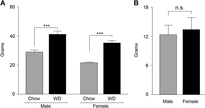
In this study, we analyzed the fatty acid profile of brains and plasma from male and female mice fed chow or a western-style high fat diet (WD) for 16 weeks to determine if males and females process fatty acids differently. Based on the differences in fatty acids observed in vivo, we performed in vitro experiments on N43 hypothalamic neuronal cells to begin to elucidate how the fatty acid milieu may impact brain inflammation.
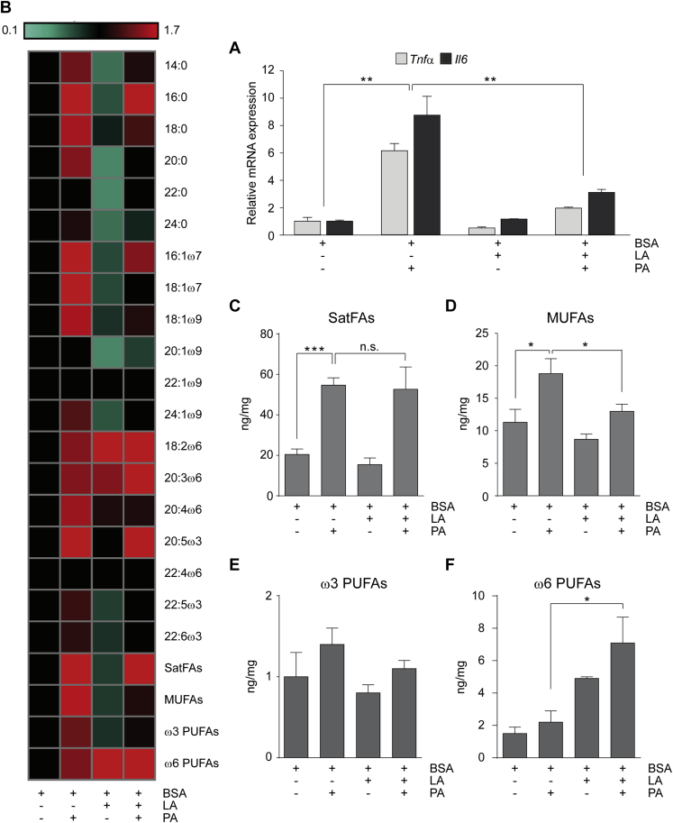
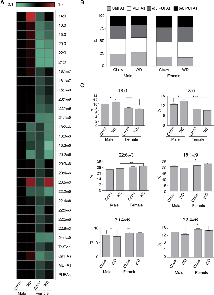
Using a comprehensive mass spectrometry fatty acid analysis, which includes a profile for 52 different fatty acid isomers, we assayed the plasma and brain fatty acid composition of age-matched male and female mice maintained on chow or a WD. Additionally, using the same techniques, we determined the fatty acid composition of N43 hypothalamic cells following exposure to palmitic and linoleic acid, alone or in combination.
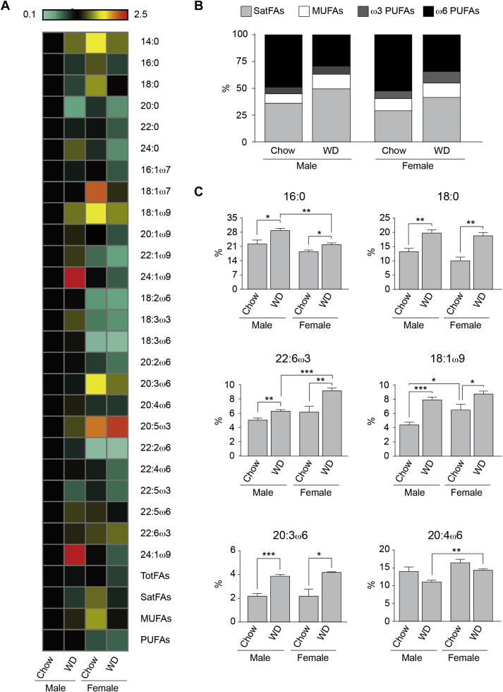
Our data demonstrate there is a sexual dimorphism in brain fatty acid content both following the consumption of the chow diet, as well as the WD, with males having an increased percentage of saturated fatty acids and reductions in ω6-polyunsaturated fatty acids when compared to females. Interestingly, we did not observe a sexual dimorphism in fatty acid content in the plasma of the same mice. Furthermore, exposure of N43 cells to the ω6-PUFA linoleic acid, which is higher in female brains when compared to males, reduces palmitic acid-induced inflammation.
Our data suggest male and female brains, and not plasma, differ in their fatty acid profile. This is the first time, to our knowledge, lipidomic analyses has been used to directly test the hypothesis there is a sexual dimorphism in brain and plasma fatty acid composition following consumption of the chow diet, as well as following exposure to the WD.
在本研究中,我们分析了喂食普通饲料或西式高脂饮食(WD)16周的雄性和雌性小鼠的大脑和血浆中的脂肪酸谱,以确定雄性和雌性处理脂肪酸的方式是否不同。基于体内观察到的脂肪酸差异,我们对N43下丘脑神经元细胞进行了体外实验,以开始阐明脂肪酸环境如何影响脑部炎症。
我们使用了一种全面的质谱脂肪酸分析方法,该方法包括52种不同脂肪酸异构体的谱图,测定了食用普通饲料或WD的年龄匹配的雄性和雌性小鼠的血浆和脑脂肪酸组成。此外,使用相同的技术,我们测定了N43下丘脑细胞在单独或联合暴露于棕榈酸和亚油酸后的脂肪酸组成。
我们的数据表明,无论是食用普通饲料还是WD后,大脑脂肪酸含量都存在性别差异,与雌性相比,雄性的饱和脂肪酸百分比增加,ω6-多不饱和脂肪酸减少。有趣的是,我们在同一小鼠的血浆脂肪酸含量中未观察到性别差异。此外,将N43细胞暴露于雌性大脑中比雄性更高的ω6-多不饱和脂肪酸亚油酸中,可减少棕榈酸诱导的炎症。
我们的数据表明,雄性和雌性的大脑而非血浆,在脂肪酸谱上存在差异。据我们所知,这是首次使用脂质组学分析直接测试以下假设:食用普通饲料以及暴露于WD后,大脑和血浆脂肪酸组成存在性别差异。