Laboratory of Clinical Cytogenetics, Department of Genetics, Yale School of MedicineNew Haven, CT, USA; Department of Pathology, Institute of Hematology and Blood Diseases Hospital, Chinese Academy of Medical SciencesTianjin, China.
Laboratory of Clinical Cytogenetics, Department of Genetics, Yale School of MedicineNew Haven, CT, USA; Department of Cell Biology and Genetics, Guangxi Medical UniversityNanning, China.
Front Cell Dev Biol. 2016 Sep 5;4:89. doi: 10.3389/fcell.2016.00089. eCollection 2016.
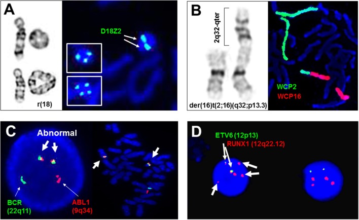
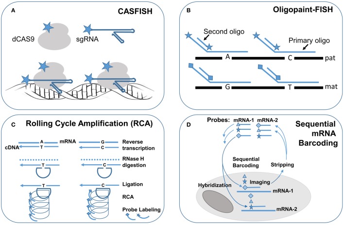
Fluorescence in situ hybridization (FISH) is a macromolecule recognition technology based on the complementary nature of DNA or DNA/RNA double strands. Selected DNA strands incorporated with fluorophore-coupled nucleotides can be used as probes to hybridize onto the complementary sequences in tested cells and tissues and then visualized through a fluorescence microscope or an imaging system. This technology was initially developed as a physical mapping tool to delineate genes within chromosomes. Its high analytical resolution to a single gene level and high sensitivity and specificity enabled an immediate application for genetic diagnosis of constitutional common aneuploidies, microdeletion/microduplication syndromes, and subtelomeric rearrangements. FISH tests using panels of gene-specific probes for somatic recurrent losses, gains, and translocations have been routinely applied for hematologic and solid tumors and are one of the fastest-growing areas in cancer diagnosis. FISH has also been used to detect infectious microbias and parasites like malaria in human blood cells. Recent advances in FISH technology involve various methods for improving probe labeling efficiency and the use of super resolution imaging systems for direct visualization of intra-nuclear chromosomal organization and profiling of RNA transcription in single cells. Cas9-mediated FISH (CASFISH) allowed in situ labeling of repetitive sequences and single-copy sequences without the disruption of nuclear genomic organization in fixed or living cells. Using oligopaint-FISH and super-resolution imaging enabled in situ visualization of chromosome haplotypes from differentially specified single-nucleotide polymorphism loci. Single molecule RNA FISH (smRNA-FISH) using combinatorial labeling or sequential barcoding by multiple round of hybridization were applied to measure mRNA expression of multiple genes within single cells. Research applications of these single molecule single cells DNA and RNA FISH techniques have visualized intra-nuclear genomic structure and sub-cellular transcriptional dynamics of many genes and revealed their functions in various biological processes.
荧光原位杂交(FISH)是一种基于 DNA 或 DNA/RNA 双链互补性的大分子识别技术。与荧光素偶联核苷酸结合的选定 DNA 链可用作探针,与测试细胞和组织中的互补序列杂交,然后通过荧光显微镜或成像系统进行可视化。该技术最初是作为一种物理图谱绘制工具开发的,用于划定染色体中的基因。其对单个基因水平的高分析分辨率以及高灵敏度和特异性使其能够立即应用于染色体结构常见非整倍体、微缺失/微重复综合征和端粒重排的遗传诊断。使用基因特异性探针组合进行体细胞反复缺失、增益和易位的 FISH 检测已常规应用于血液学和实体肿瘤,是癌症诊断中增长最快的领域之一。FISH 还用于检测人类血液中的感染性微生物和寄生虫,如疟疾。FISH 技术的最新进展涉及各种提高探针标记效率的方法,以及使用超分辨率成像系统直接可视化核内染色体组织和单个细胞中 RNA 转录的分析。Cas9 介导的 FISH(CASFISH)允许在固定或活细胞中不破坏核基因组组织的情况下对重复序列和单拷贝序列进行原位标记。使用寡核苷酸-FISH 和超分辨率成像可以在不同的单核苷酸多态性位点原位可视化染色体单倍型。使用组合标记或通过多轮杂交进行的顺序条形码的单分子 RNA FISH(smRNA-FISH)用于测量单个细胞内多个基因的 mRNA 表达。这些单分子单细胞 DNA 和 RNA FISH 技术的研究应用可视化了核内基因组结构和许多基因的亚细胞转录动力学,并揭示了它们在各种生物过程中的功能。