Boult Jessica K R, Borri Marco, Jury Alexa, Popov Sergey, Box Gary, Perryman Lara, Eccles Suzanne A, Jones Chris, Robinson Simon P
Division of Radiotherapy and Imaging, The Institute of Cancer Research, London, UK.
Royal Marsden NHS Foundation Trust, Sutton, Surrey, UK.
NMR Biomed. 2016 Nov;29(11):1608-1617. doi: 10.1002/nbm.3594. Epub 2016 Sep 27.
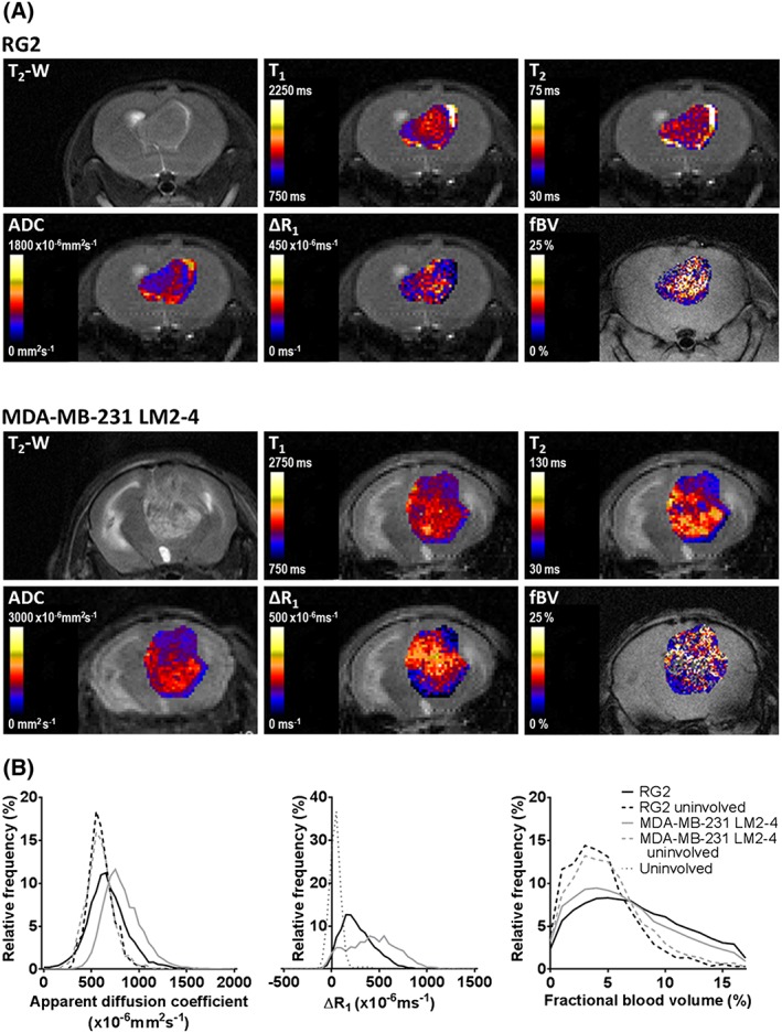
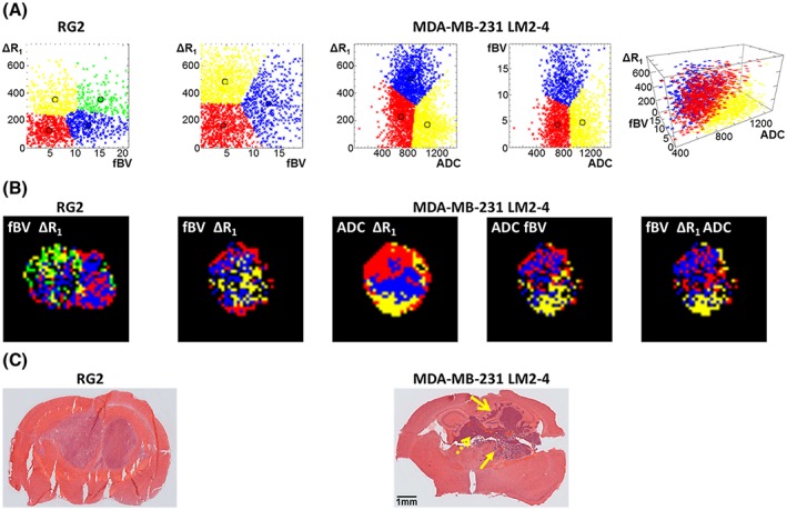
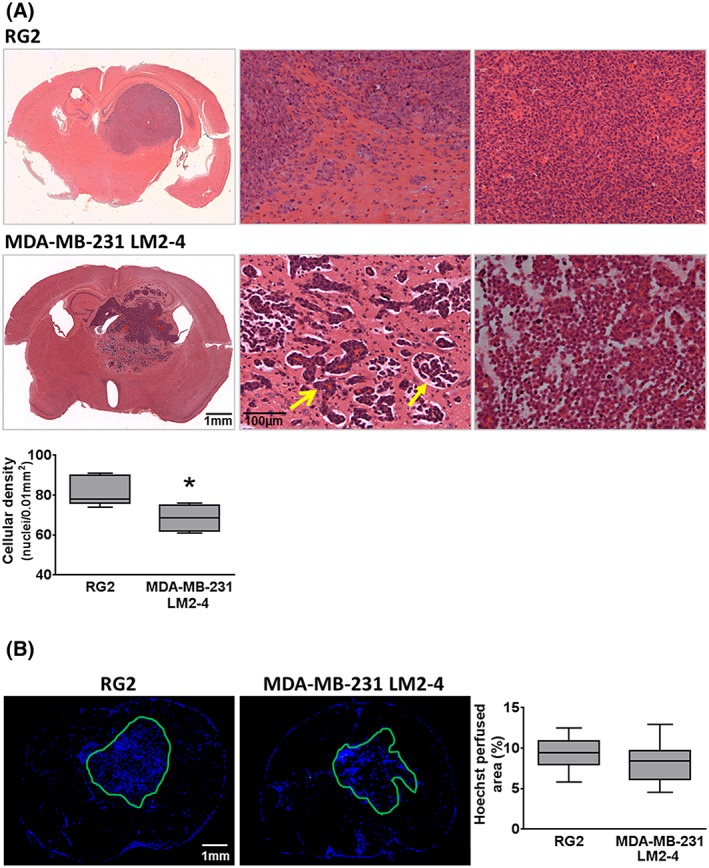
High grade and metastatic brain tumours exhibit considerable spatial variations in proliferation, angiogenesis, invasion, necrosis and oedema. Vascular heterogeneity arising from vascular co-option in regions of invasive growth (in which the blood-brain barrier remains intact) and neoangiogenesis is a major challenge faced in the assessment of brain tumours by conventional MRI. A multiparametric MRI approach, incorporating native measurements and both Gd-DTPA (Magnevist) and ultrasmall superparamagnetic iron oxide (P904)-enhanced imaging, was used in combination with histogram and unsupervised cluster analysis using a k-means algorithm to examine the spatial distribution of vascular parameters, water diffusion characteristics and invasion in intracranially propagated rat RG2 gliomas and human MDA-MB-231 LM2-4 breast adenocarcinomas in mice. Both tumour models presented with higher ΔR (the change in transverse relaxation rate R induced by Gd-DTPA), fractional blood volume (fBV) and apparent diffusion coefficient than uninvolved regions of the brain. MDA-MB-231 LM2-4 tumours were less densely cellular than RG2 tumours and exhibited substantial local invasion, associated with oedema, whereas invasion in RG2 tumours was minimal. These additional features were reflected in the more heterogeneous appearance of MDA-MB-231 LM2-4 tumours on T -weighted images and maps of functional MRI parameters. Unsupervised cluster analysis separated subregions with distinct functional properties; areas with a low fBV and relatively impermeable blood vessels (low ΔR ) were predominantly located at the tumour margins, regions of MDA-MB-231 LM2-4 tumours with relatively high levels of water diffusion and low vascular permeability and/or fBV corresponded to histologically identified regions of invasion and oedema, and areas of mismatch between vascular permeability and blood volume were identified. We demonstrate that dual contrast MRI and evaluation of tissue diffusion properties, coupled with cluster analysis, allows for the assessment of heterogeneity within invasive brain tumours and the designation of functionally diverse subregions that may provide more informative predictive biomarkers.
高级别和转移性脑肿瘤在增殖、血管生成、侵袭、坏死和水肿方面表现出显著的空间差异。在侵袭性生长区域(血脑屏障保持完整)中由血管共选和新生血管生成引起的血管异质性是传统MRI评估脑肿瘤时面临的主要挑战。采用多参数MRI方法,结合原始测量以及钆喷酸葡胺(马根维显)和超小超顺磁性氧化铁(P904)增强成像,并使用k均值算法进行直方图分析和无监督聚类分析,以研究颅内传播的大鼠RG2胶质瘤和小鼠人MDA-MB-231 LM2-4乳腺腺癌中血管参数、水扩散特征和侵袭的空间分布。两种肿瘤模型的横向弛豫率变化(ΔR,由钆喷酸葡胺诱导的横向弛豫率R的变化)、血容量分数(fBV)和表观扩散系数均高于脑内未受累区域。MDA-MB-231 LM2-4肿瘤的细胞密度低于RG2肿瘤,并表现出大量局部侵袭,伴有水肿,而RG2肿瘤中的侵袭极少。这些额外特征反映在T加权图像和功能MRI参数图上MDA-MB-231 LM2-4肿瘤更不均匀的外观上。无监督聚类分析分离出具有不同功能特性的子区域;fBV低且血管相对不通透(ΔR低)的区域主要位于肿瘤边缘,MDA-MB-231 LM2-4肿瘤中具有相对高水平水扩散和低血管通透性和/或fBV的区域对应于组织学确定的侵袭和水肿区域,并且识别出了血管通透性和血容量不匹配的区域。我们证明,双对比MRI和组织扩散特性评估,结合聚类分析,能够评估侵袭性脑肿瘤内 的异质性,并确定功能多样的子区域,这些子区域可能提供更具信息性的预测生物标志物。