Tymofiyeva Olga, Connolly Colm G, Ho Tiffany C, Sacchet Matthew D, Henje Blom Eva, LeWinn Kaja Z, Xu Duan, Yang Tony T
Department of Radiology and Biomedical Imaging, University of California San Francisco, United States.
Department of Psychiatry, University of California San Francisco, United States.
J Affect Disord. 2017 Jan 1;207:18-25. doi: 10.1016/j.jad.2016.09.013. Epub 2016 Sep 19.
Adolescence is a vulnerable period for the onset of major depressive disorder (MDD). While some studies have shown white matter alterations in adolescent MDD, there is still a gap in understanding how the brain is affected at a network level.
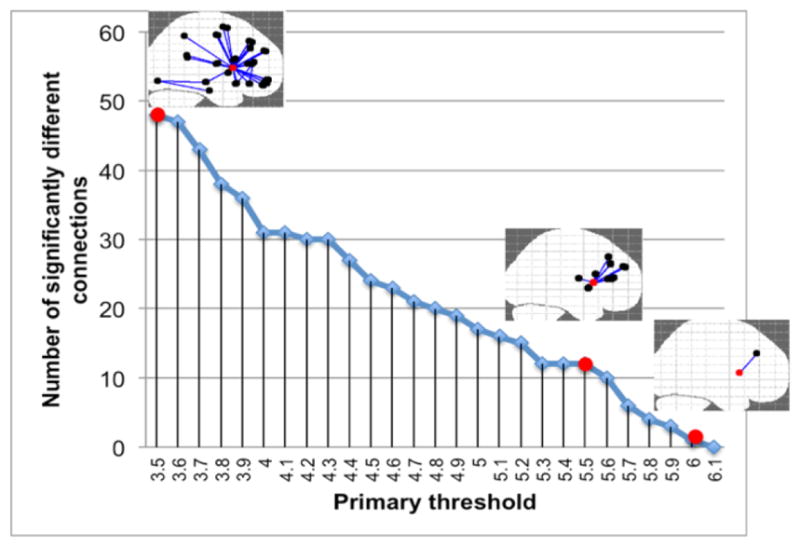
We compared diffusion tensor imaging (DTI)-based brain networks in a cohort of 57 adolescents with MDD and 41 well-matched healthy controls who completed self-reports of depression symptoms and stressful life events. Using atlas-based brain regions as network nodes and tractography streamline count or mean fractional anisotropy (FA) as edge weights, we examined weighted local and global network properties and performed Network-Based Statistic (NBS) analysis.
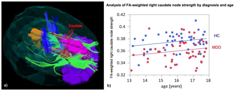
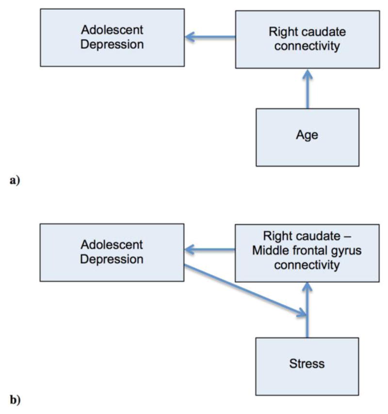
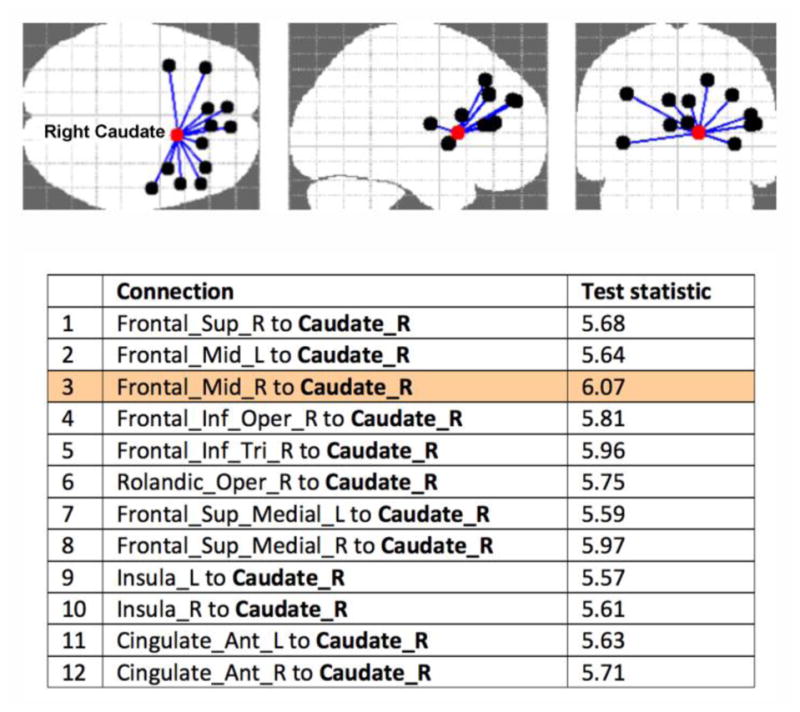
While there were no significant group differences in the global network properties, the FA-weighted node strength of the right caudate was significantly lower in depressed adolescents and correlated positively with age across both groups. The NBS analysis revealed a cluster of lower FA-based connectivity in depressed subjects centered on the right caudate, including connections to frontal gyri, insula, and anterior cingulate. Within this cluster, the most robust difference between groups was the connection between the right caudate and middle frontal gyrus. This connection showed a significant diagnosis by stress interaction and a negative correlation with total stress in depressed adolescents.
Use of DTI-based tractography, one atlas-based parcellation, and FA values to characterize brain networks represent this study's limitations.
Our results allowed us to suggest caudate-centric models of dysfunctional processes underlying adolescent depression, which might guide future studies and help better understand and treat this disorder.
青春期是重度抑郁症(MDD)发病的脆弱时期。虽然一些研究已经表明青少年MDD存在白质改变,但在理解大脑在网络层面如何受到影响方面仍存在差距。
我们比较了57名患有MDD的青少年和41名匹配良好的健康对照者基于扩散张量成像(DTI)的脑网络,这些参与者完成了抑郁症状和应激性生活事件的自我报告。以基于图谱的脑区作为网络节点,以纤维束成像流线计数或平均分数各向异性(FA)作为边权重,我们检查了加权局部和全局网络属性,并进行了基于网络的统计(NBS)分析。
虽然全局网络属性在两组之间没有显著差异,但抑郁青少年右侧尾状核的FA加权节点强度显著较低,且在两组中均与年龄呈正相关。NBS分析显示,抑郁受试者中以右侧尾状核为中心存在一组基于FA的连通性较低的区域,包括与额回、岛叶和前扣带回的连接。在这个区域内,两组之间最显著的差异是右侧尾状核与额中回之间的连接。这种连接在抑郁青少年中显示出应激交互作用的显著诊断价值,并且与总应激呈负相关。
使用基于DTI的纤维束成像、一种基于图谱的脑区划分以及FA值来表征脑网络是本研究的局限性。
我们的结果使我们能够提出以尾状核为中心的青少年抑郁症潜在功能失调过程模型,这可能为未来的研究提供指导,并有助于更好地理解和治疗这种疾病。