• 文献检索
  • 文档翻译
  • 深度研究
  • 学术资讯
  • Suppr Zotero 插件Zotero 插件
  • 邀请有礼
  • 套餐&价格
  • 历史记录
应用&插件
Suppr Zotero 插件Zotero 插件浏览器插件Mac 客户端Windows 客户端微信小程序
定价
高级版会员购买积分包购买API积分包
服务
文献检索文档翻译深度研究API 文档MCP 服务
关于我们
关于 Suppr公司介绍联系我们用户协议隐私条款
关注我们

Suppr 超能文献

核心技术专利:CN118964589B侵权必究
粤ICP备2023148730 号-1Suppr @ 2026

文献检索

告别复杂PubMed语法,用中文像聊天一样搜索,搜遍4000万医学文献。AI智能推荐,让科研检索更轻松。

立即免费搜索

文件翻译

保留排版,准确专业,支持PDF/Word/PPT等文件格式,支持 12+语言互译。

免费翻译文档

深度研究

AI帮你快速写综述,25分钟生成高质量综述,智能提取关键信息,辅助科研写作。

立即免费体验

非增强计算机断层扫描在脑静脉窦血栓形成诊断中的应用

Non-contrast computed tomography in the diagnosis of cerebral venous sinus thrombosis.

作者信息

Avsenik Jernej, Oblak Janja Pretnar, Popovic Katarina Surlan

机构信息

Clinical Institute of Radiology, University Medical Centre, Ljubljana, Slovenia.

Department of Neurology, University Medical Centre, Ljubljana, Slovenia.

出版信息

Radiol Oncol. 2016 Jul 19;50(3):263-8. doi: 10.1515/raon-2016-0026. eCollection 2016 Sep 1.

DOI:10.1515/raon-2016-0026
PMID:27679541
原文链接:https://pmc.ncbi.nlm.nih.gov/articles/PMC5024657/
Abstract

BACKGROUND

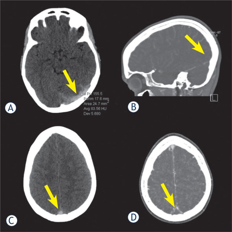
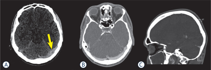
The aim of the study was to investigate the sensitivity and specificity of non-contrast computed tomography (NCCT) in the diagnosis of cerebral venous sinus thrombosis (CVST). Methods. Screening our neurological department database, we identified 53 patients who were admitted to neurological emergency department with clinical signs of CVST. Two independent observers assessed the NCCT scans for the presence of CVST. CT venography and/or MR venography were used as a reference standard. Interobserver agreement between the two readers was assessed using Kappa statistic. Attenuation inside the cerebral venous sinuses was measured and compared between the patient and the control group.

RESULTS

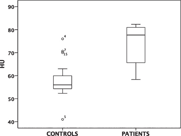
CVST was confirmed in 13 patients. Sensitivity and specificity of NCCT for overall presence of CVST were 100% and 83%, respectively, with Kappa value of 0.72 (a good agreement between observers). The attenuation values between CVST patients and control group were significantly different (73.4 ± 14.12 HU vs. 58.1 ± 7.58 HU; p = 0.000). The ROC analysis showed an area under the curve (AUC) of 0.916 (95% CI, 0.827 - 1.00) and an optimal cutoff value of 64 HU, leading to a sensitivity of 85% and specificity of 87%.

CONCLUSIONS

NCCT as a first-line investigation has a high value for diagnosis of CVST in the emergency setting. The additional measurement of the sinus attenuation may improve the diagnostic value of the examination.

摘要

背景

本研究旨在探讨非增强计算机断层扫描(NCCT)在诊断脑静脉窦血栓形成(CVST)中的敏感性和特异性。方法:通过筛查我们神经科数据库,我们确定了53例因CVST临床症状入住神经急诊科的患者。两名独立观察者评估NCCT扫描是否存在CVST。CT静脉造影和/或MR静脉造影用作参考标准。使用Kappa统计量评估两位读者之间的观察者间一致性。测量并比较患者和对照组脑静脉窦内的衰减。

结果

13例患者确诊为CVST。NCCT对CVST总体存在情况的敏感性和特异性分别为100%和83%,Kappa值为0.72(观察者之间一致性良好)。CVST患者与对照组之间的衰减值有显著差异(73.4±14.12 HU对58.1±7.58 HU;p = 0.000)。ROC分析显示曲线下面积(AUC)为0.916(95%CI,0.827 - 1.00),最佳截断值为64 HU,敏感性为85%,特异性为87%。

结论

在紧急情况下,NCCT作为一线检查对CVST的诊断具有很高价值。额外测量窦内衰减可能会提高检查的诊断价值。

https://cdn.ncbi.nlm.nih.gov/pmc/blobs/2ee0/5024657/58ef9cba0fa2/j_raon-2016-0026_fig_003.jpg
https://cdn.ncbi.nlm.nih.gov/pmc/blobs/2ee0/5024657/6b5c8747f5a8/j_raon-2016-0026_fig_001.jpg
https://cdn.ncbi.nlm.nih.gov/pmc/blobs/2ee0/5024657/5b1834d7271c/j_raon-2016-0026_fig_002.jpg
https://cdn.ncbi.nlm.nih.gov/pmc/blobs/2ee0/5024657/58ef9cba0fa2/j_raon-2016-0026_fig_003.jpg
https://cdn.ncbi.nlm.nih.gov/pmc/blobs/2ee0/5024657/6b5c8747f5a8/j_raon-2016-0026_fig_001.jpg
https://cdn.ncbi.nlm.nih.gov/pmc/blobs/2ee0/5024657/5b1834d7271c/j_raon-2016-0026_fig_002.jpg
https://cdn.ncbi.nlm.nih.gov/pmc/blobs/2ee0/5024657/58ef9cba0fa2/j_raon-2016-0026_fig_003.jpg

相似文献

1
Non-contrast computed tomography in the diagnosis of cerebral venous sinus thrombosis.非增强计算机断层扫描在脑静脉窦血栓形成诊断中的应用
Radiol Oncol. 2016 Jul 19;50(3):263-8. doi: 10.1515/raon-2016-0026. eCollection 2016 Sep 1.
2
Utility of Hounsfield unit and hematocrit values in the diagnosis of acute venous sinus thrombosis in unenhanced brain CTs in the pediatric population.亨氏单位和血细胞比容值在儿科人群未增强脑部CT诊断急性静脉窦血栓形成中的应用
Pediatr Radiol. 2019 Feb;49(2):234-239. doi: 10.1007/s00247-018-4273-y. Epub 2018 Oct 16.
3
Diagnostic Accuracy of Hounsfield Unit Value and Hounsfield Unit to Hematocrit Ratio in Predicting Cerebral Venous Sinus Thrombosis: A Retrospective Case-Control Study.亨氏单位值及亨氏单位与血细胞比容比值在预测脑静脉窦血栓形成中的诊断准确性:一项回顾性病例对照研究
Cureus. 2024 Apr 3;16(4):e57567. doi: 10.7759/cureus.57567. eCollection 2024 Apr.
4
Plain CT vs MR venography in acute cerebral venous sinus thrombosis: Triumphant dark horse.急性脑静脉窦血栓形成中平扫CT与磁共振静脉血管造影:脱颖而出的黑马。
Indian J Radiol Imaging. 2018 Jul-Sep;28(3):280-284. doi: 10.4103/ijri.IJRI_328_17.
5
Dural venous sinus thrombosis: The combination of noncontrast CT, MRI and PC-MR venography to enhance accuracy.硬脑膜静脉窦血栓形成:非增强CT、MRI和PC-MR静脉造影相结合以提高准确性。
Neuroradiol J. 2018 Oct;31(5):473-481. doi: 10.1177/1971400918781969. Epub 2018 Jun 5.
6
Are cerebral veins hounsfield unit and H: H ratio calculating in unenhanced CT eligible to diagnosis of acute cerebral vein thrombosis?在未增强CT中计算脑静脉的亨氏单位和H:H比值是否适用于急性脑静脉血栓形成的诊断?
J Res Med Sci. 2019 Sep 30;24:83. doi: 10.4103/jrms.JRMS_1027_18. eCollection 2019.
7
Diagnostic value of non-contrast brain computed tomography in the evaluation of acute cerebral venous thrombosis.非增强脑计算机断层扫描在急性脑静脉血栓形成评估中的诊断价值。
Sci Rep. 2020 Jan 21;10(1):883. doi: 10.1038/s41598-020-57867-1.
8
Wavelet-Based Angiographic Reconstruction of Computed Tomography Perfusion Data: Diagnostic Value in Cerebral Venous Sinus Thrombosis.基于小波的计算机断层扫描灌注数据血管造影重建:对脑静脉窦血栓形成的诊断价值
Invest Radiol. 2017 May;52(5):302-309. doi: 10.1097/RLI.0000000000000337.
9
Radiological evaluation in patients with clinical suspicion of cerebral venous sinus thrombosis presenting with nontraumatic headache - a retrospective observational study with a validation cohort.临床怀疑脑静脉窦血栓形成并伴有非创伤性头痛患者的放射学评估 - 一项回顾性观察研究及验证队列。
BMC Med Imaging. 2020 Feb 27;20(1):24. doi: 10.1186/s12880-020-00426-x.
10
Quantitative non-contrast measurements improve diagnosing dural venous sinus thrombosis.定量非增强测量有助于诊断硬脑膜静脉窦血栓形成。
Neuroradiology. 2016 Jul;58(7):657-63. doi: 10.1007/s00234-016-1681-2. Epub 2016 Apr 4.

引用本文的文献

1
Dual- energy CT versus single-energy CT for estimation of hematocrit and hemoglobin in the brain: an in vivo analysis.双能CT与单能CT用于估计脑内血细胞比容和血红蛋白的比较:一项活体分析
Neuroradiology. 2025 Jul 14. doi: 10.1007/s00234-025-03700-3.
2
Attenuation measurements on non-contrast-enhanced CT brain: a re-visit of their role in the diagnosis of cerebral venous thrombosis.非增强CT脑衰减测量:重新审视其在脑静脉血栓形成诊断中的作用。
Jpn J Radiol. 2025 Jun;43(6):940-948. doi: 10.1007/s11604-025-01746-0. Epub 2025 Feb 18.
3
Determining the Diagnostic Value of Venous Sinus Density Indices in Non-Contrast Brain CT Scan for Early Diagnosis of Cerebral Venous Sinus Thrombosis.

本文引用的文献

1
CT density measurement and H:H ratio are useful in diagnosing acute cerebral venous sinus thrombosis.CT 密度测量和 H:H 比值有助于诊断急性脑静脉窦血栓形成。
AJNR Am J Neuroradiol. 2013 Aug;34(8):1568-72. doi: 10.3174/ajnr.A3469. Epub 2013 Mar 7.
2
Cerebral venous thrombosis.脑静脉血栓形成
Circulation. 2012 Apr 3;125(13):1704-9. doi: 10.1161/CIRCULATIONAHA.111.067835.
3
Acute abdominal venous thromboses--the hyperdense CT sign.急性腹部静脉血栓形成——CT高密度征
确定非增强脑部CT扫描中静脉窦密度指数对脑静脉窦血栓形成早期诊断的诊断价值。
Brain Behav. 2025 Feb;15(2):e70324. doi: 10.1002/brb3.70324.
4
Overview of the utility of noncontrast CT brain in the diagnosis of cerebral vein thrombosis.非增强头颅CT在脑静脉血栓形成诊断中的应用概述
Radiol Case Rep. 2024 Nov 13;20(1):736-742. doi: 10.1016/j.radcr.2024.10.035. eCollection 2025 Jan.
5
Post-Traumatic Cerebral Venous Sinus Thrombosis (PtCVST) Resulting in Increased Intracranial Pressure during Early Post-Traumatic Brain Injury Period: Case Report and Narrative Literature Review.创伤性脑损伤早期发生创伤后脑静脉窦血栓形成(PtCVST)导致颅内压升高:病例报告及文献综述
Healthcare (Basel). 2024 Sep 1;12(17):1743. doi: 10.3390/healthcare12171743.
6
Diagnostic and Therapeutic Challenges of Concurrent Intracranial Hemorrhage and Cerebral Venous Thrombosis in a Patient With Acute Lymphoblastic Leukemia: A Case Report and Literature Review.急性淋巴细胞白血病患者并发颅内出血和脑静脉血栓形成的诊断与治疗挑战:一例报告及文献综述
Cureus. 2023 Apr 12;15(4):e37482. doi: 10.7759/cureus.37482. eCollection 2023 Apr.
7
Diagnostic Sensitivity of Unenhanced CT for Cerebral Venous Thrombosis: Can Clot Density Measurement Replace CT Venogram?未增强CT对脑静脉血栓形成的诊断敏感性:血栓密度测量能否取代CT静脉造影?
Indian J Radiol Imaging. 2023 Feb 11;33(2):187-194. doi: 10.1055/s-0043-1761184. eCollection 2023 Apr.
8
Cerebral Venous Thrombosis: A Challenging Diagnosis; A New Nonenhanced Computed Tomography Standardized Semi-Quantitative Method.脑静脉血栓形成:诊断难题;一种新的非增强 CT 标准化半定量方法。
Tomography. 2021 Dec 22;8(1):1-9. doi: 10.3390/tomography8010001.
9
Acute cerebral venous thrombosis - still an underdiagnosed pathology in emergency computed tomography of the brain.急性脑静脉血栓形成——在脑部急诊计算机断层扫描中仍是一种诊断不足的病症。
Pol J Radiol. 2021 Oct 8;86:e574-e582. doi: 10.5114/pjr.2021.109490. eCollection 2021.
10
COVID-19 vaccine-induced immune thrombotic thrombocytopenia: A review of the potential mechanisms and proposed management.COVID-19 疫苗诱导的免疫性血栓性血小板减少症:潜在机制的综述及提出的管理建议。
Sci Prog. 2021 Apr-Jun;104(2):368504211025927. doi: 10.1177/00368504211025927.
J Comput Assist Tomogr. 2012 Jan-Feb;36(1):8-13. doi: 10.1097/RCT.0b013e3182436c86.
4
Cerebral venous sinus density on noncontrast CT correlates with hematocrit.非增强 CT 上脑静脉窦密度与红细胞压积相关。
AJNR Am J Neuroradiol. 2011 Aug;32(7):1354-7. doi: 10.3174/ajnr.A2504. Epub 2011 May 12.
5
Diagnosis and management of cerebral venous thrombosis: a statement for healthcare professionals from the American Heart Association/American Stroke Association.脑静脉血栓形成的诊断和治疗:美国心脏协会/美国中风协会向医疗保健专业人员的声明。
Stroke. 2011 Apr;42(4):1158-92. doi: 10.1161/STR.0b013e31820a8364. Epub 2011 Feb 3.
6
Noncontrast CT in deep cerebral venous thrombosis and sinus thrombosis: comparison of its diagnostic value for both entities.非增强CT在大脑深部静脉血栓形成和静脉窦血栓形成中的应用:对两者诊断价值的比较
AJNR Am J Neuroradiol. 2009 Apr;30(4):728-35. doi: 10.3174/ajnr.A1451. Epub 2009 Feb 12.
7
Isolated lateral sinus thrombosis: a series of 62 patients.孤立性横窦血栓形成:62例患者系列研究
Stroke. 2009 Feb;40(2):476-81. doi: 10.1161/STROKEAHA.107.509711. Epub 2008 Dec 31.
8
Diagnostic role of 64-slice multidetector row CT scan and CT venogram in cases of cerebral venous thrombosis.64层多排螺旋CT扫描及CT静脉成像在脑静脉血栓形成病例中的诊断作用
Emerg Radiol. 2008 Sep;15(5):325-33. doi: 10.1007/s10140-008-0723-4. Epub 2008 Apr 24.
9
Risk factors of cerebral vein and sinus thrombosis.脑静脉和静脉窦血栓形成的危险因素。
Front Neurol Neurosci. 2008;23:23-54. doi: 10.1159/000111259.
10
Cerebral venous thrombosis: an update.脑静脉血栓形成:最新进展
Lancet Neurol. 2007 Feb;6(2):162-70. doi: 10.1016/S1474-4422(07)70029-7.