Baban Babak, Liu Jun Yao, Payne Samuel, Abebe Worku, Yu Jack C, Mozaffari Mahmood S
Department of Oral Biology; CL-2140, Dental College of Georgia, Augusta University, Augusta, GA 30912-1128 USA ; Department of Surgery, Section of Plastic Surgery, Medical College of Georgia, Augusta, GA 30912 USA.
Department of Oral Biology; CL-2140, Dental College of Georgia, Augusta University, Augusta, GA 30912-1128 USA.
EPMA J. 2016 Oct 4;7(1):21. doi: 10.1186/s13167-016-0070-6. eCollection 2016.
Recruitment of stem cells to sites of tissue injury constitutes an important mechanism aimed at tissue repair and regeneration. However, it is not clear how the diabetic milieu affects the viability of endogenous stem cells. Thus, we tested the hypothesis that diabetes mellitus is associated with increased apoptosis which, in turn, contributes to reduction in stem cells and the manifestation of type 2 diabetic nephropathy.
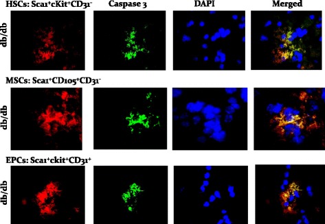
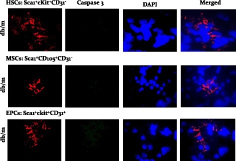
Sixteen-week-old male obese type 2 diabetic db/db mice, and their appropriate controls, were used for assessment of the status of endothelial progenitor cells (EPCs), mesenchymal stem cells (MSCs), and hematopoetic stem cells (HSCs) in the peripheral blood and renal tissue using specific cell markers. Further, we explored whether diabetic animals display greater apoptosis of stem cell subsets.
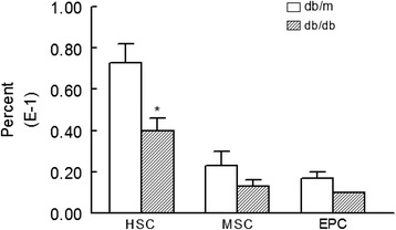
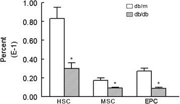
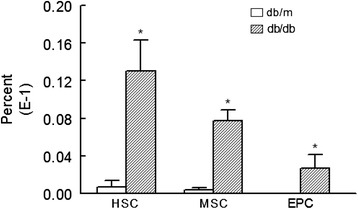
The peripheral blood cells of db/db mice displayed reduction in EPCs ( < 0.05) compared to those of db/m controls. Further, kidney cells prepared from experimental groups also showed reductions in EPCs, MSCs, and HSCs. We also observed increased apoptosis of stem cell subsets in cells prepared from kidneys of db/db than those of db/m mice.
The present study shows a similar pattern of decline in stem cell subsets in peripheral blood and kidneys of db/db mice, an effect likely related to increased apoptosis. Collectively, the results suggest that apoptosis of stem cells likely contributes to eventual manifestation of renal failure in diabetes mellitus. Monitoring of blood levels of stem cell subsets could predict failure of their reparative and protective effects and eventual manifestations of diabetic complications.
将干细胞募集到组织损伤部位是组织修复和再生的重要机制。然而,糖尿病环境如何影响内源性干细胞的活力尚不清楚。因此,我们验证了以下假设:糖尿病与细胞凋亡增加相关,而细胞凋亡反过来又会导致干细胞数量减少以及2型糖尿病肾病的表现。
使用16周龄雄性肥胖2型糖尿病db/db小鼠及其相应对照,通过特异性细胞标志物评估外周血和肾组织中内皮祖细胞(EPC)、间充质干细胞(MSC)和造血干细胞(HSC)的状态。此外,我们探究了糖尿病动物的干细胞亚群是否表现出更大程度的凋亡。
与db/m对照小鼠相比,db/db小鼠的外周血细胞中EPC数量减少(P<0.05)。此外,实验组制备的肾细胞中EPC、MSC和HSC也减少。我们还观察到,与db/m小鼠相比,db/db小鼠肾脏制备的细胞中干细胞亚群的凋亡增加。
本研究显示db/db小鼠外周血和肾脏中干细胞亚群呈现相似的减少模式,这种效应可能与细胞凋亡增加有关。总体而言,结果表明干细胞凋亡可能导致糖尿病肾病中肾衰竭的最终表现。监测干细胞亚群的血液水平可以预测其修复和保护作用的失效以及糖尿病并发症的最终表现。