• 文献检索
  • 文档翻译
  • 深度研究
  • 学术资讯
  • Suppr Zotero 插件Zotero 插件
  • 邀请有礼
  • 套餐&价格
  • 历史记录
应用&插件
Suppr Zotero 插件Zotero 插件浏览器插件Mac 客户端Windows 客户端微信小程序
定价
高级版会员购买积分包购买API积分包
服务
文献检索文档翻译深度研究API 文档MCP 服务
关于我们
关于 Suppr公司介绍联系我们用户协议隐私条款
关注我们

Suppr 超能文献

核心技术专利:CN118964589B侵权必究
粤ICP备2023148730 号-1Suppr @ 2026

文献检索

告别复杂PubMed语法,用中文像聊天一样搜索,搜遍4000万医学文献。AI智能推荐,让科研检索更轻松。

立即免费搜索

文件翻译

保留排版,准确专业,支持PDF/Word/PPT等文件格式,支持 12+语言互译。

免费翻译文档

深度研究

AI帮你快速写综述,25分钟生成高质量综述,智能提取关键信息,辅助科研写作。

立即免费体验

磁共振胰胆管成像和内镜超声诊断胰腺分裂症的准确性:系统评价和荟萃分析。

The Accuracies of Diagnosing Pancreas Divisum by Magnetic Resonance Cholangiopancreatography and Endoscopic Ultrasound: A Systematic Review and Meta-analysis.

机构信息

Department of Gastroenterology, The First Affiliated Hospital, College of Medicine, Zhejiang University, 310003 Hangzhou, China.

Molecular Hepatology-Alcohol Associated Diseases, II. Medical Clinic Faculty of Medicine at Mannheim, University of Heidelberg, 68167 Mannheim, Germany.

出版信息

Sci Rep. 2016 Oct 13;6:35389. doi: 10.1038/srep35389.

DOI:10.1038/srep35389
PMID:27734952
原文链接:https://pmc.ncbi.nlm.nih.gov/articles/PMC5062127/
Abstract

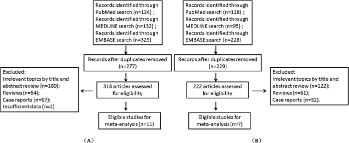
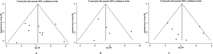
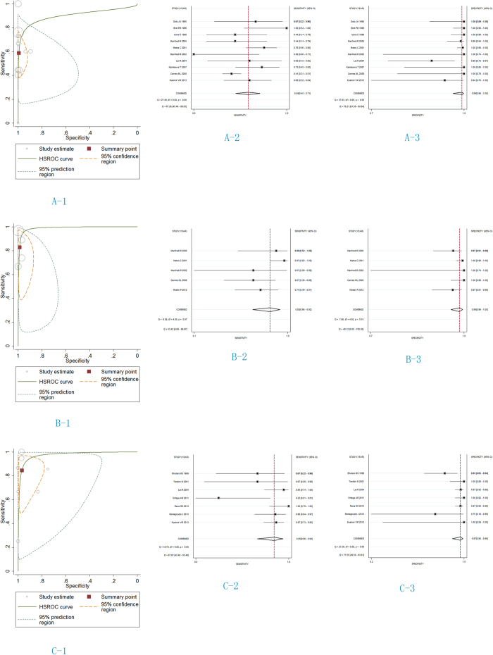
Magnetic resonance cholangiopancreatography (MRCP), MRCP after secretin stimulation (S-MRCP) and endoscopic ultrasonography (EUS) are all selected to diagnose pancreas divisum. However, the accuracies of three diagnosis remain unclear. The aim is to address the diagnostic accuracies of MRCP, S-MRCP and EUS on pancreas divisum. We searched PubMed, MEDLINE and EMBASE databases from inception to January, 2015. Of the 536 citations retrieved, 16 studies were included. For MRCP diagnosis on pancreas divisum, the area under the hierarchical summary receiver-operating characteristic (HSROC) curve was 0.90 (95% confidence interval [CI] 0.87 to 0.92), and for S-MRCP and EUS, 0.99 (95% CI 0.97 to 0.99) and 0.97 (95% CI 0.96 to 0.98). Sensitivity and specificity for MRCP were 0.59 (95% CI 0.45 to 0.71) and 0.99 (95% CI 0.96 to 1.00); for S-MRCP, 0.83 (95% CI 0.66 to 0.92) and 0.99 (95% CI 0.96 to 1.00); for EUS, 0.85 (95% CI 0.67 to 0.94) and 0.97 (95% CI 0.90 to 0.99). Comprehensive comparison of three diagnostic techniques to pancreas divisum, S-MRCP was more reliable than MRCP and EUS on the effect of the diagnostic test.

摘要

磁共振胆胰管成像(MRCP)、促胰液素刺激后的 MRCP(S-MRCP)和内镜超声检查(EUS)均被用于诊断胰腺分裂症。然而,三种诊断方法的准确性尚不清楚。本研究旨在评估 MRCP、S-MRCP 和 EUS 对胰腺分裂症的诊断准确性。我们检索了从建库至 2015 年 1 月的 PubMed、MEDLINE 和 EMBASE 数据库。从 536 条引文中共纳入了 16 项研究。MRCP 诊断胰腺分裂症的综合受试者工作特征(HSROC)曲线下面积为 0.90(95%置信区间 [CI] 0.87 至 0.92),S-MRCP 和 EUS 的曲线下面积分别为 0.99(95% CI 0.97 至 0.99)和 0.97(95% CI 0.96 至 0.98)。MRCP 的敏感度和特异度分别为 0.59(95% CI 0.45 至 0.71)和 0.99(95% CI 0.96 至 1.00);S-MRCP 为 0.83(95% CI 0.66 至 0.92)和 0.99(95% CI 0.96 至 1.00);EUS 为 0.85(95% CI 0.67 至 0.94)和 0.97(95% CI 0.90 至 0.99)。综合比较三种诊断技术,S-MRCP 对胰腺分裂症的诊断效果优于 MRCP 和 EUS。

https://cdn.ncbi.nlm.nih.gov/pmc/blobs/d7c3/5062127/18928162aa28/srep35389-f3.jpg
https://cdn.ncbi.nlm.nih.gov/pmc/blobs/d7c3/5062127/6fc5b13b692b/srep35389-f1.jpg
https://cdn.ncbi.nlm.nih.gov/pmc/blobs/d7c3/5062127/d548f67bffd5/srep35389-f2.jpg
https://cdn.ncbi.nlm.nih.gov/pmc/blobs/d7c3/5062127/18928162aa28/srep35389-f3.jpg
https://cdn.ncbi.nlm.nih.gov/pmc/blobs/d7c3/5062127/6fc5b13b692b/srep35389-f1.jpg
https://cdn.ncbi.nlm.nih.gov/pmc/blobs/d7c3/5062127/d548f67bffd5/srep35389-f2.jpg
https://cdn.ncbi.nlm.nih.gov/pmc/blobs/d7c3/5062127/18928162aa28/srep35389-f3.jpg

相似文献

1
The Accuracies of Diagnosing Pancreas Divisum by Magnetic Resonance Cholangiopancreatography and Endoscopic Ultrasound: A Systematic Review and Meta-analysis.磁共振胰胆管成像和内镜超声诊断胰腺分裂症的准确性:系统评价和荟萃分析。
Sci Rep. 2016 Oct 13;6:35389. doi: 10.1038/srep35389.
2
Comparison of EUS with MRCP in idiopathic acute pancreatitis: a systematic review and meta-analysis.超声内镜与磁共振胰胆管成像在特发性急性胰腺炎中的比较:系统评价和荟萃分析。
Gastrointest Endosc. 2018 May;87(5):1180-1188.e9. doi: 10.1016/j.gie.2017.11.028. Epub 2017 Dec 7.
3
Imaging modalities for characterising focal pancreatic lesions.用于表征胰腺局灶性病变的成像方式。
Cochrane Database Syst Rev. 2017 Apr 17;4(4):CD010213. doi: 10.1002/14651858.CD010213.pub2.
4
Transabdominal ultrasound and endoscopic ultrasound for diagnosis of gallbladder polyps.经腹超声和内镜超声用于胆囊息肉的诊断。
Cochrane Database Syst Rev. 2018 Aug 15;8(8):CD012233. doi: 10.1002/14651858.CD012233.pub2.
5
A systematic review on endoscopic detection rate, endotherapy, and surgery for pancreas divisum.关于胰腺分裂症的内镜检出率、内镜治疗及手术治疗的系统评价
Endoscopy. 2009 May;41(5):439-44. doi: 10.1055/s-0029-1214505. Epub 2009 Mar 31.
6
Prospective comparison of endoscopic ultrasonography and magnetic resonance cholangiopancreatography in the etiological diagnosis of "idiopathic" acute pancreatitis.内镜超声检查与磁共振胰胆管成像在“特发性”急性胰腺炎病因诊断中的前瞻性比较。
Pancreas. 2011 Mar;40(2):289-94. doi: 10.1097/MPA.0b013e318201654a.
7
Accuracy of magnetic resonance cholangiopancreatography in the diagnosis of pancreas divisum.磁共振胆胰管成像诊断胰腺分裂症的准确性。
Dig Dis Sci. 2012 Jan;57(1):170-4. doi: 10.1007/s10620-011-1823-7. Epub 2011 Jul 15.
8
Sensitivity of endoscopic ultrasound, multidetector computed tomography, and magnetic resonance cholangiopancreatography in the diagnosis of pancreas divisum: a tertiary center experience.内镜超声、多排螺旋 CT 和磁共振胰胆管成像在胰腺分裂症诊断中的敏感性:一家三级中心的经验。
Pancreas. 2013 Apr;42(3):436-41. doi: 10.1097/MPA.0b013e31826c711a.
9
Pancreatico-biliary endoscopic ultrasound: a systematic review of the levels of evidence, performance and outcomes.胰腺胆道内镜超声:证据水平、性能和结果的系统评价。
World J Gastroenterol. 2012 Aug 28;18(32):4243-56. doi: 10.3748/wjg.v18.i32.4243.
10
Magnetic resonance cholangiopancreatography in the diagnosis of pancreas divisum: a systematic review and meta-analysis.磁共振胰胆管造影术在胰腺分裂症诊断中的应用:一项系统评价和荟萃分析。
Pancreas. 2014 Aug;43(6):823-8. doi: 10.1097/MPA.0000000000000143.

引用本文的文献

1
Pancreas Divisum With Santorinicele: A Rare Cause of Recurrent Acute Pancreatitis.胰腺分裂伴桑托里尼囊肿:复发性急性胰腺炎的罕见病因。
Cureus. 2025 Apr 18;17(4):e82498. doi: 10.7759/cureus.82498. eCollection 2025 Apr.
2
Early Chronic Pancreatitis Findings by Endoscopic Ultrasonography (EUS) in Asymptomatic Patients with Pancreas Divisum.内镜超声检查(EUS)对胰腺分裂无症状患者早期慢性胰腺炎的发现
Diagnostics (Basel). 2025 Jan 22;15(3):253. doi: 10.3390/diagnostics15030253.
3
Pancreas divisum unmasked: a case report.胰腺分裂症的发现:一例病例报告

本文引用的文献

1
Endoscopic approach to the diagnosis and treatment of pancreatic disease.内镜技术在胰腺疾病诊断与治疗中的应用
Curr Opin Gastroenterol. 2014 Sep;30(5):524-30. doi: 10.1097/MOG.0000000000000095.
2
Role of endoscopic ultrasound in the diagnosis of pancreas divisum.内镜超声在胰腺分裂症诊断中的作用。
Endosc Ultrasound. 2013 Jan;2(1):7-10. doi: 10.7178/eus.04.003.
3
Sensitivity of endoscopic ultrasound, multidetector computed tomography, and magnetic resonance cholangiopancreatography in the diagnosis of pancreas divisum: a tertiary center experience.
Pan Afr Med J. 2024 Oct 22;49:48. doi: 10.11604/pamj.2024.49.48.45004. eCollection 2024.
4
Refractory postoperative pancreatic fistula following laparoscopic distal pancreatectomy for pancreatic cancer caused by incomplete pancreas divisum: a case report.腹腔镜胰体尾切除术治疗胰腺癌术后难治性胰瘘:不完全胰腺分裂症一例报告。
Clin J Gastroenterol. 2024 Jun;17(3):587-591. doi: 10.1007/s12328-024-01942-x. Epub 2024 Mar 9.
5
Endoscopic Ultrasound in Pancreatic Duct Anomalies.内镜超声在胰管异常中的应用
Diagnostics (Basel). 2023 Oct 5;13(19):3129. doi: 10.3390/diagnostics13193129.
6
Pancreas divisum causing recurrent pancreatitis in a young patient: A case report.胰腺分裂症致青年患者复发性胰腺炎一例报告
Radiol Case Rep. 2023 Jul 28;18(10):3535-3538. doi: 10.1016/j.radcr.2023.07.028. eCollection 2023 Oct.
7
Magnetic resonance cholangiopancreatography enhanced by virtual reality as a novel tool to improve the understanding of biliary anatomy and the teaching of surgical trainees.虚拟现实增强磁共振胰胆管造影术作为一种新型工具,可提高对胆道解剖结构的理解以及对外科实习生的教学水平。
Front Surg. 2022 Aug 12;9:916443. doi: 10.3389/fsurg.2022.916443. eCollection 2022.
8
Endoscopic ultrasound diagnostic gain over computed tomography and magnetic resonance cholangiopancreatography in defining etiology of idiopathic acute pancreatitis.在明确特发性急性胰腺炎病因方面,内镜超声相对于计算机断层扫描和磁共振胰胆管造影的诊断优势。
World J Gastrointest Endosc. 2022 Jun 16;14(6):0. doi: 10.4253/wjge.v14.i6.0000.
9
Identification and management of pancreas divisum.胰腺分体的识别与处理。
Expert Rev Gastroenterol Hepatol. 2019 Nov;13(11):1089-1105. doi: 10.1080/17474124.2019.1685871. Epub 2019 Nov 8.
10
Acute Pancreatitis in Pancreas Divisum Secondary to an Impacted Stone in the Minor Papilla.胰腺分裂症继发小乳头结石嵌顿所致的急性胰腺炎。
Cureus. 2019 Aug 25;11(8):e5481. doi: 10.7759/cureus.5481.
内镜超声、多排螺旋 CT 和磁共振胰胆管成像在胰腺分裂症诊断中的敏感性:一家三级中心的经验。
Pancreas. 2013 Apr;42(3):436-41. doi: 10.1097/MPA.0b013e31826c711a.
4
Accuracy of magnetic resonance cholangiopancreatography in the diagnosis of pancreas divisum.磁共振胆胰管成像诊断胰腺分裂症的准确性。
Dig Dis Sci. 2012 Jan;57(1):170-4. doi: 10.1007/s10620-011-1823-7. Epub 2011 Jul 15.
5
Risk factors for ERCP-related complications in patients with pancreas divisum: a retrospective study.胰腺分裂患者行内镜逆行胰胆管造影相关并发症的危险因素:一项回顾性研究。
Gastrointest Endosc. 2011 May;73(5):963-70. doi: 10.1016/j.gie.2010.12.035. Epub 2011 Mar 9.
6
Prospective comparison of endoscopic ultrasonography and magnetic resonance cholangiopancreatography in the etiological diagnosis of "idiopathic" acute pancreatitis.内镜超声检查与磁共振胰胆管成像在“特发性”急性胰腺炎病因诊断中的前瞻性比较。
Pancreas. 2011 Mar;40(2):289-94. doi: 10.1097/MPA.0b013e318201654a.
7
Diagnostic yield and reliability of endoscopic ultrasonography in patients with idiopathic acute pancreatitis.内镜超声检查在特发性急性胰腺炎患者中的诊断率及可靠性
Scand J Gastroenterol. 2010 Mar;45(3):375-81. doi: 10.3109/00365520903508894.
8
Miss rate of pancreas divisum by magnetic resonance cholangiopancreatography in clinical practice.临床实践中磁共振胰胆管造影对胰腺分裂症的漏诊率
Pancreas. 2008 Aug;37(2):151-3. doi: 10.1097/MPA.0b013e318164cbaf.
9
Hierarchical models for ROC curve summary measures: design and analysis of multi-reader, multi-modality studies of medical tests.用于ROC曲线汇总指标的分层模型:医学检验多阅片者、多模态研究的设计与分析
Stat Med. 2008 Jan 30;27(2):243-56. doi: 10.1002/sim.2828.
10
Bivariate meta-analysis of sensitivity and specificity with sparse data: a generalized linear mixed model approach.稀疏数据下灵敏度和特异度的双变量Meta分析:一种广义线性混合模型方法
J Clin Epidemiol. 2006 Dec;59(12):1331-2; author reply 1332-3. doi: 10.1016/j.jclinepi.2006.06.011. Epub 2006 Sep 28.