Canavese Federico, Dimeglio Alain, Barbetta Davide, Galeotti Marco, Canavese Bartolomeo, Cavalli Fabio
Department of Pediatric Surgery, University Hospital Estaing, 1 Place Lucie et Raymond Aubrac, 63003 Clermont-Ferrand, France "University of Montpellier, 34000 Montpellier, France.
Department of Pediatric Orthopedic Surgery, Saint Roch Hospital, 8 rue Marguerite, France; University of Montpellier, Faculty of Medicine, 2 Rue de l'Ecole de Medecine, 34000 Montpellier, France.
Indian J Orthop. 2016 Sep;50(5):558-566. doi: 10.4103/0019-5413.189600.
This experimental study provides a qualitative description and the morpho-structural features of the fusions taking place in the thoracic spine between prepubertal age and skeletal maturity. There is a lack of informations regarding the influence of partial or total dorso-thoracic vertebral arthrodesis on the development of the thoracic cage as well as its potential effects on different intra and extra-thoracic organs. This study admits the hypothesis that vertebral arthrodesis may have influence on other body areas and so, it intends to verify the possible secondary involvement of other body parts, such as intervertebral discs, cervical and thoracic spinal ganglia, sternocostal cartilage, ovaries and lungs.
Fifty-four female New Zealand white rabbits were submitted to dorsal arthrodesis. The radiologic imaging and light microscopy histological pictures were taken and studied in all. Computed tomography (CT) scan measurements were performed in operated and sham operated rabbits at different time. Similarly, histological specimens of intervertebral discs, cervical and thoracic spinal ganglia, sternocostal cartilage, ovaries and lungs were analyzed at different times. The study ended at the age of 17-18 months.
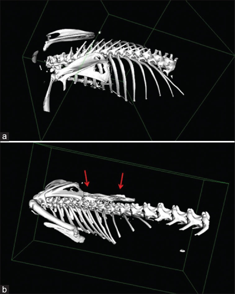
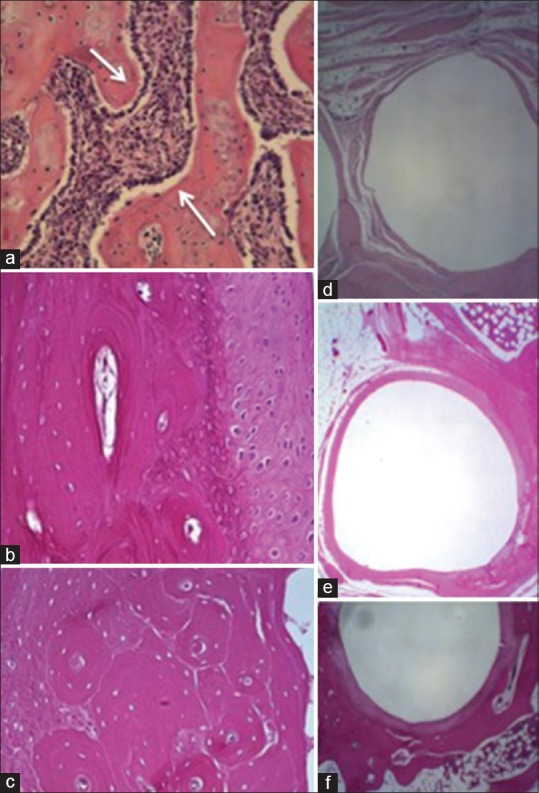
Most rabbits had formed a fusion mass, which was only fibrous at first, then osteofibrous and finally, in the older subjects, structured in lamellar-osteon tissue. Intervertebral foramens were negatively involved in vertebral arthrodesis, as shown by CT scans. Intervertebral discs showed irregular aspects. The increase of atresic follicles and the reduction of primordial follicles in operated rabbits led to the hypothesis of a cause-effect relationship between arthrodesis and modified hormonal status. Dorsal root ganglia showed microscopic alterations in operated rabbits especially.
The process of fusion mass and bone formation, associated with the arthrodesis, involves at different degrees of the vertebral bodies, discs and intervertebral foramens, ganglia and spinal nerve roots.
本实验研究对青春期前至骨骼成熟阶段胸椎融合的形态结构特征进行了定性描述。目前缺乏关于部分或全胸段椎体融合术对胸廓发育的影响及其对胸内、外不同器官潜在作用的信息。本研究提出一个假设,即椎体融合术可能会影响身体其他部位,因此旨在验证其他身体部位如椎间盘、颈胸段脊神经节、胸肋软骨、卵巢和肺是否可能受到继发性影响。
对54只雌性新西兰白兔进行背部融合术。对所有兔子进行放射影像学检查并拍摄光学显微镜组织学图片。在不同时间对手术组和假手术组兔子进行计算机断层扫描(CT)测量。同样,在不同时间对椎间盘、颈胸段脊神经节、胸肋软骨、卵巢和肺的组织学标本进行分析。研究在17 - 18个月龄时结束。
大多数兔子形成了融合块,最初仅为纤维性,然后是骨纤维性,最终在年龄较大的兔子中形成板层骨组织。CT扫描显示椎间孔在椎体融合术中受到负面影响。椎间盘呈现不规则形态。手术组兔子闭锁卵泡增加而原始卵泡减少,这引发了融合术与激素状态改变之间存在因果关系的假设。尤其在手术组兔子中,背根神经节出现了微观改变。
与融合术相关的融合块形成和骨形成过程,在不同程度上涉及椎体、椎间盘、椎间孔、神经节和脊神经根。