Yang Tian, Wang Jinyuan, Pang Yamei, Dang Xiaomin, Ren Hui, Liu Ya, Chen Mingwei, Shang Dong
Department of Respiratory and Critical Care Medicine, First Affiliated Hospital of Xi'an Jiaotong University, Xi'an, Shaanxi 710061, P.R. China.
Department of Clinical Laboratory, First Affiliated Hospital of Xi'an Jiaotong University, Xi'an, Shaanxi 710061, P.R. China.
Mol Med Rep. 2016 Nov;14(5):4643-4649. doi: 10.3892/mmr.2016.5838. Epub 2016 Oct 12.
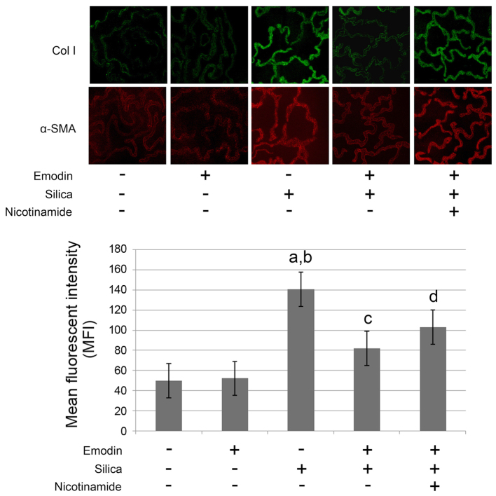
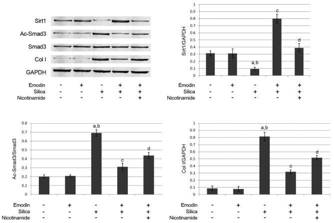
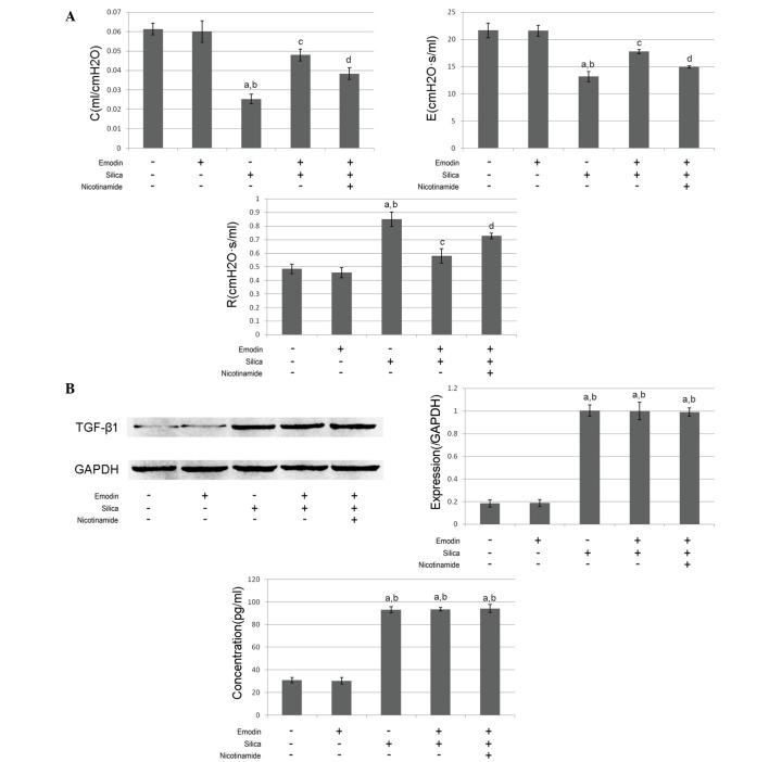
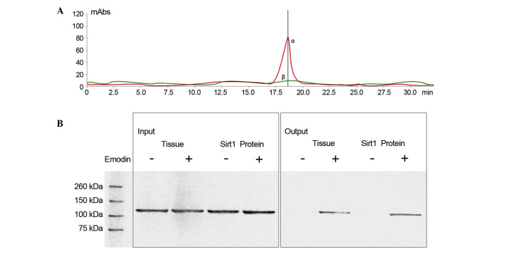
Pulmonary silicosis is characterized by lung fibrosis, which leads to impairment of pulmonary function; the specific mechanism remains to be fully elucidated Emodin shows antifibrotic effects in several organs with fibrosis, however, it has not been investigated in pulmonary silicosis. In the present study, the possible mechanism of lung fibrosis and the antifibrotic effect of emodin in silica inhalation‑induced lung fibrosis were investigated. Pulmonary silica particle inhalation was used to induce lung fibrosis in mice. Emodin and or the sirtuin 1 (Sirt1) inhibitor, nicotinamide, were used to treat the modeled animals. Pulmonary function was assessed using an occlusion method. The deposition of collagen I and α‑smooth muscle actin (SMA) in the lung tissue were detected using fluorescence staining; transforming growth factor‑β1 (TGF‑β1) in the bronchoalveolar lavage fluid (BALF) was examined using an enzyme‑linked immunosorbent assay; TGF-β1/Sirt1/small mothers against decapentaplegic (Smad) signaling activation in lung tissue was also examined. The molecular contacts between emodin were evaluated using liquid chromatography‑mass spectrometry analysis. The deposition of collagen I and α‑SMA in lung tissues were found to be elevated following silica exposure, however, this was relieved by emodin treatment. The pulmonary function of the animals was impaired by silica inhalation, and this was improved by emodin administration. However, the therapeutic effects of emodin on lung fibrosis were impaired by nicotinamide administration. The levels of TGF‑β1 in the BALF and lung tissue were elevated by silica inhalation, however, they were not affected by either emodin or nicotinamide treatment. Additionally, emodin was found to increase the expression level of Sirt1, which decreased the level of deacetylated Smad3 to attenuate collagen deposition. Furthermore, the data suggested that there was direct binding between emodin and Sirt1. Sirt1‑regulated TGF‑β1/Smad signaling was involved in silica inhalation‑induced lung fibrosis. Emodin attenuated this lung fibrosis to improve pulmonary function by targeting Sirt1, which regulated TGF-β1/Smad fibrotic signaling.
肺硅沉着病以肺纤维化为特征,可导致肺功能受损;其具体机制仍有待充分阐明。大黄素在几种纤维化器官中显示出抗纤维化作用,然而,尚未对其在肺硅沉着病中的作用进行研究。在本研究中,对肺纤维化的可能机制以及大黄素在二氧化硅吸入诱导的肺纤维化中的抗纤维化作用进行了研究。采用吸入肺二氧化硅颗粒的方法诱导小鼠肺纤维化。使用大黄素和/或沉默调节蛋白1(Sirt1)抑制剂烟酰胺对模型动物进行治疗。采用阻断法评估肺功能。使用荧光染色检测肺组织中I型胶原蛋白和α-平滑肌肌动蛋白(SMA)的沉积;采用酶联免疫吸附测定法检测支气管肺泡灌洗液(BALF)中转化生长因子-β1(TGF-β1);还检测了肺组织中TGF-β1/Sirt1/抗五聚体蛋白母系小蛋白(Smad)信号激活情况。使用液相色谱-质谱分析法评估大黄素之间的分子相互作用。发现二氧化硅暴露后肺组织中I型胶原蛋白和α-SMA的沉积增加,然而,大黄素治疗可缓解这种情况。吸入二氧化硅会损害动物的肺功能,而给予大黄素可改善肺功能。然而,给予烟酰胺会削弱大黄素对肺纤维化的治疗效果。吸入二氧化硅会使BALF和肺组织中TGF-β1的水平升高,然而,大黄素或烟酰胺治疗对其没有影响。此外,发现大黄素可增加Sirt1的表达水平,从而降低去乙酰化Smad3的水平,以减少胶原蛋白沉积。此外,数据表明大黄素与Sirt1之间存在直接结合。Sirt1调节的TGF-β1/Smad信号通路参与了二氧化硅吸入诱导的肺纤维化。大黄素通过靶向Sirt1来减轻这种肺纤维化,从而改善肺功能,Sirt1调节TGF-β1/Smad纤维化信号通路。