• 文献检索
  • 文档翻译
  • 深度研究
  • 学术资讯
  • Suppr Zotero 插件Zotero 插件
  • 邀请有礼
  • 套餐&价格
  • 历史记录
应用&插件
Suppr Zotero 插件Zotero 插件浏览器插件Mac 客户端Windows 客户端微信小程序
定价
高级版会员购买积分包购买API积分包
服务
文献检索文档翻译深度研究API 文档MCP 服务
关于我们
关于 Suppr公司介绍联系我们用户协议隐私条款
关注我们

Suppr 超能文献

核心技术专利:CN118964589B侵权必究
粤ICP备2023148730 号-1Suppr @ 2026

文献检索

告别复杂PubMed语法,用中文像聊天一样搜索,搜遍4000万医学文献。AI智能推荐,让科研检索更轻松。

立即免费搜索

文件翻译

保留排版,准确专业,支持PDF/Word/PPT等文件格式,支持 12+语言互译。

免费翻译文档

深度研究

AI帮你快速写综述,25分钟生成高质量综述,智能提取关键信息,辅助科研写作。

立即免费体验

上斜肌麻痹中的眼外肌间隙

Extraocular Muscle Compartments in Superior Oblique Palsy.

作者信息

Suh Soh Youn, Clark Robert A, Le Alan, Demer Joseph L

机构信息

Department of Ophthalmology, David Geffen Medical School at University of California, Los Angeles, Los Angeles, California.

Department of Ophthalmology, David Geffen Medical School at University of California, Los Angeles, Los Angeles, California 2David Geffen Medical School at University of California, Los Angeles, Los Angeles, California.

出版信息

Invest Ophthalmol Vis Sci. 2016 Oct 1;57(13):5535-5540. doi: 10.1167/iovs.16-20172.

DOI:10.1167/iovs.16-20172
PMID:27768791
原文链接:https://pmc.ncbi.nlm.nih.gov/articles/PMC5080936/
Abstract

PURPOSE

To investigate changes in volumes of extraocular muscle (EOM) compartments in unilateral superior oblique (SO) palsy using magnetic resonance imaging (MRI).

METHODS

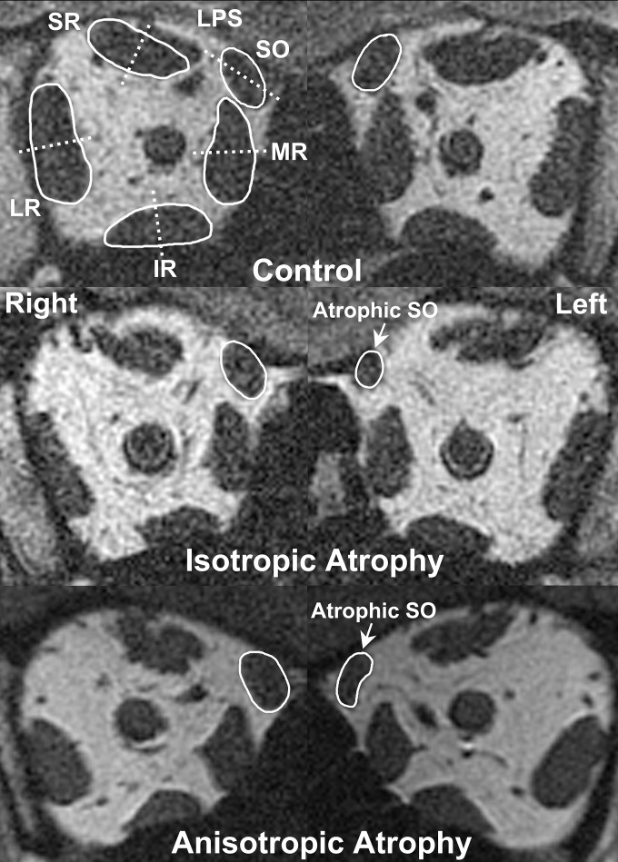
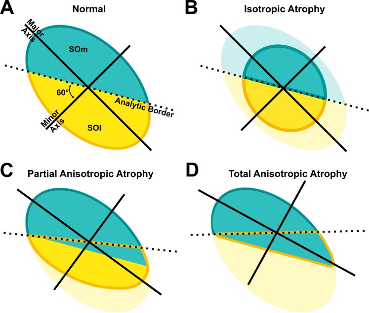
High-resolution, surface-coil MRI was obtained in 19 patients with unilateral SO palsy and 19 age-matched orthotropic control subjects. Rectus EOMs and the SO were divided into two anatomic compartments for volume analysis in patients with unilateral SO palsy, allowing comparison of total compartmental volumes versus controls. Medial and lateral compartmental volumes of the SO muscle were compared in patients with isotropic (round shape) versus anisotropic (elongated shape) SO atrophy.

RESULTS

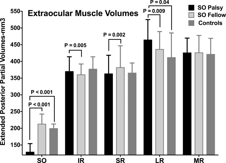
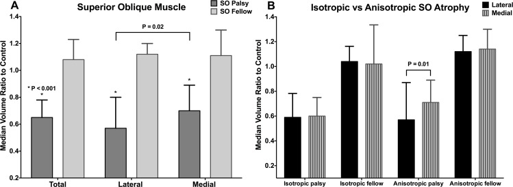
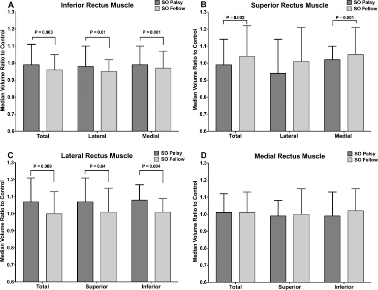
The medial and lateral compartments of the ipsilesional SO muscles were equally atrophic in isotropic SO palsy, whereas the lateral compartment was significantly smaller than the medial in anisotropic SO palsy (P = 0.01). In contrast to the SO, there were no differential compartmental volume changes in rectus EOMs; however, there was significant total muscle hypertrophy in the ipsilesional inferior rectus (IR) and lateral rectus (LR) muscles and contralesional superior rectus (SR) muscles. Medial rectus (MR) volume was normal both ipsi- and contralesionally.

CONCLUSIONS

A subset of patients with SO palsy exhibit selective atrophy of the lateral, predominantly vertically acting SO compartment. Superior oblique atrophy is associated with whole-muscle volume changes in the ipsilesional IR, ipsilesional LR, and contralesional SR; however, SO muscle atrophy is not associated with compartmentally selective volume changes in the rectus EOMs. Selective compartmental SO pathology may provide an anatomic mechanism that explains some of the variability in clinical presentations of SO palsy.

摘要

目的

使用磁共振成像(MRI)研究单侧上斜肌(SO)麻痹患者眼外肌(EOM)各部分的体积变化。

方法

对19例单侧SO麻痹患者和19例年龄匹配的正视对照者进行高分辨率表面线圈MRI检查。在单侧SO麻痹患者中,将直肌EOM和SO分为两个解剖部分进行体积分析,以便与对照组比较各部分总体积。比较等轴状(圆形)与非等轴状(细长形)SO萎缩患者的SO肌内侧和外侧部分体积。

结果

在等轴状SO麻痹中,患侧SO肌的内侧和外侧部分萎缩程度相同,而在非等轴状SO麻痹中,外侧部分明显小于内侧部分(P = 0.01)。与SO不同,直肌EOM各部分体积没有差异变化;然而,患侧下直肌(IR)和外直肌(LR)以及对侧上直肌(SR)出现明显的整体肌肉肥大。内直肌(MR)同侧和对侧的体积均正常。

结论

一部分SO麻痹患者表现出外侧主要垂直作用的SO部分选择性萎缩。上斜肌萎缩与患侧IR、患侧LR和对侧SR的全肌体积变化有关;然而,SO肌萎缩与直肌EOM各部分选择性体积变化无关。SO部分的选择性病理改变可能提供一种解剖学机制,解释SO麻痹临床表现的一些变异性。

https://cdn.ncbi.nlm.nih.gov/pmc/blobs/3c12/5080936/cd4c863c84a8/i1552-5783-57-13-5535-f05.jpg
https://cdn.ncbi.nlm.nih.gov/pmc/blobs/3c12/5080936/dd1d0f667cc5/i1552-5783-57-13-5535-f01.jpg
https://cdn.ncbi.nlm.nih.gov/pmc/blobs/3c12/5080936/c732e6a380ff/i1552-5783-57-13-5535-f02.jpg
https://cdn.ncbi.nlm.nih.gov/pmc/blobs/3c12/5080936/3575e1495e77/i1552-5783-57-13-5535-f03.jpg
https://cdn.ncbi.nlm.nih.gov/pmc/blobs/3c12/5080936/822dd11c8e12/i1552-5783-57-13-5535-f04.jpg
https://cdn.ncbi.nlm.nih.gov/pmc/blobs/3c12/5080936/cd4c863c84a8/i1552-5783-57-13-5535-f05.jpg

相似文献

1
Extraocular Muscle Compartments in Superior Oblique Palsy.上斜肌麻痹中的眼外肌间隙
Invest Ophthalmol Vis Sci. 2016 Oct 1;57(13):5535-5540. doi: 10.1167/iovs.16-20172.
2
Rectus Pulley Displacements without Abnormal Oblique Contractility Explain Strabismus in Superior Oblique Palsy.无异常斜肌收缩的直肌滑车移位可解释上斜肌麻痹性斜视。
Ophthalmology. 2016 Jun;123(6):1222-31. doi: 10.1016/j.ophtha.2016.02.016. Epub 2016 Mar 13.
3
Enhanced vertical rectus contractility by magnetic resonance imaging in superior oblique palsy.磁共振成像显示上斜肌麻痹时垂直直肌收缩力增强
Arch Ophthalmol. 2011 Jul;129(7):904-8. doi: 10.1001/archophthalmol.2011.152.
4
Magnetic resonance imaging of the functional anatomy of the inferior oblique muscle in superior oblique palsy.上斜肌麻痹时下斜肌功能解剖的磁共振成像
Ophthalmology. 2003 Jun;110(6):1219-29. doi: 10.1016/S0161-6420(03)00331-2.
5
Magnetic resonance imaging of bilateral split lateral rectus transposition to the medial globe.双侧外直肌劈开移位至眼球内侧的磁共振成像
Graefes Arch Clin Exp Ophthalmol. 2015 Sep;253(9):1587-90. doi: 10.1007/s00417-015-3071-6. Epub 2015 Jul 2.
6
Magnetic resonance imaging of the functional anatomy of the inferior rectus muscle in superior oblique muscle palsy.上斜肌麻痹时下直肌功能解剖的磁共振成像
Ophthalmology. 2008 Nov;115(11):2079-86. doi: 10.1016/j.ophtha.2008.04.040. Epub 2008 Aug 9.
7
Orbital magnetic resonance imaging of extraocular muscles in chronic progressive external ophthalmoplegia: specific diagnostic findings.慢性进行性眼外肌麻痹患者眼外肌的眼眶磁共振成像:特异性诊断结果
J AAPOS. 2006 Oct;10(5):414-8. doi: 10.1016/j.jaapos.2006.04.012.
8
Expanding repertoire in the oculomotor periphery: selective compartmental function in rectus extraocular muscles.在眼运动神经外周扩大曲目:直肌外眼肌的选择性隔室功能。
Ann N Y Acad Sci. 2011 Sep;1233:8-16. doi: 10.1111/j.1749-6632.2011.06112.x.
9
Functional morphometry demonstrates extraocular muscle compartmental contraction during vertical gaze changes.功能形态测量学显示,在垂直注视变化过程中眼外肌出现节段性收缩。
J Neurophysiol. 2016 Jan 1;115(1):370-8. doi: 10.1152/jn.00825.2015. Epub 2015 Nov 4.
10
Superior oblique extraocular muscle shape in superior oblique palsy.上斜肌麻痹时上斜肌的眼外肌形态
Am J Ophthalmol. 2015 Jun;159(6):1169-1179.e2. doi: 10.1016/j.ajo.2015.02.019. Epub 2015 Mar 4.

引用本文的文献

1
Functional Anatomy of Ocular Counter-Rolling in Superior Oblique Palsy.上斜肌麻痹时眼球反向转动的功能解剖学
Invest Ophthalmol Vis Sci. 2025 Sep 2;66(12):6. doi: 10.1167/iovs.66.12.6.
2
Can Binocular Alignment Distinguish Hypertropia in Sagging Eye Syndrome From Superior Oblique Palsy?双眼视功能检查能否鉴别重症肌无力性上睑下垂与上斜肌麻痹所致的斜视?
Invest Ophthalmol Vis Sci. 2022 Sep 1;63(10):13. doi: 10.1167/iovs.63.10.13.
3
Masquerading Superior Oblique Palsy.伪装性上斜肌麻痹。

本文引用的文献

1
Functional morphometry demonstrates extraocular muscle compartmental contraction during vertical gaze changes.功能形态测量学显示,在垂直注视变化过程中眼外肌出现节段性收缩。
J Neurophysiol. 2016 Jan 1;115(1):370-8. doi: 10.1152/jn.00825.2015. Epub 2015 Nov 4.
2
Compartmental Innervation of the Superior Oblique Muscle in Mammals.哺乳动物上斜肌的分区神经支配
Invest Ophthalmol Vis Sci. 2015 Oct;56(11):6237-46. doi: 10.1167/iovs.15-17602.
3
Superior oblique extraocular muscle shape in superior oblique palsy.上斜肌麻痹时上斜肌的眼外肌形态
Am J Ophthalmol. 2022 Oct;242:197-208. doi: 10.1016/j.ajo.2022.05.017. Epub 2022 May 23.
4
Normative measurements of the superior oblique and inferior oblique muscles by magnetic resonance imaging.通过磁共振成像对上斜肌和下斜肌进行规范性测量。
Surg Radiol Anat. 2022 Apr;44(4):521-525. doi: 10.1007/s00276-022-02915-w. Epub 2022 Mar 8.
5
The oblique extraocular muscles in cetaceans: Overall architecture and accessory insertions.鲸目动物的眼外斜肌:整体结构和副起点。
J Anat. 2021 Apr;238(4):917-941. doi: 10.1111/joa.13347. Epub 2020 Oct 31.
6
Translation and eccentric rotation in ocular motor modeling.眼球运动建模中的翻译和偏旋。
Prog Brain Res. 2019;248:117-126. doi: 10.1016/bs.pbr.2019.04.036. Epub 2019 May 23.
7
Functional anatomy of extraocular muscles during human vergence compensation of horizontal heterophoria.人眼水平隐斜视聚散补偿时眼外肌的功能解剖
J Neurophysiol. 2019 Jul 1;122(1):105-117. doi: 10.1152/jn.00152.2019. Epub 2019 May 1.
8
Magnetic Resonance Imaging of the Globe-Tendon Interface for Extraocular Muscles: Is There an "Arc of Contact"?眼球外肌肌腱-球结合部的磁共振成像:是否存在“接触弧”?
Am J Ophthalmol. 2018 Oct;194:170-181. doi: 10.1016/j.ajo.2018.07.002. Epub 2018 Jul 19.
Am J Ophthalmol. 2015 Jun;159(6):1169-1179.e2. doi: 10.1016/j.ajo.2015.02.019. Epub 2015 Mar 4.
4
Magnetic resonance imaging demonstrates compartmental muscle mechanisms of human vertical fusional vergence.磁共振成像显示了人类垂直融合性聚散的眼外肌机制。
J Neurophysiol. 2015 Apr 1;113(7):2150-63. doi: 10.1152/jn.00871.2014. Epub 2015 Jan 14.
5
Magnetic resonance imaging of differential compartmental function of horizontal rectus extraocular muscles during conjugate and converged ocular adduction.水平直肌在共轭性和集合性眼球内收过程中不同肌间隔功能的磁共振成像
J Neurophysiol. 2014 Aug 15;112(4):845-55. doi: 10.1152/jn.00649.2013. Epub 2014 May 21.
6
Lateral rectus superior compartment palsy.上直肌外侧肌间隙麻痹。
Am J Ophthalmol. 2014 Feb;157(2):479-487.e2. doi: 10.1016/j.ajo.2013.09.027. Epub 2013 Sep 29.
7
Selective activation of intra-muscular compartments within the trapezius muscle in subjects with Subacromial Impingement Syndrome. A case-control study.肩胛带撞击综合征患者斜方肌内肌间室的选择性激活。一项病例对照研究。
J Electromyogr Kinesiol. 2014 Feb;24(1):58-64. doi: 10.1016/j.jelekin.2013.09.008. Epub 2013 Oct 16.
8
Evaluation of the neuromuscular compartments in the peroneus longus muscle through electrical stimulation and accelerometry.通过电刺激和加速计评估腓骨长肌的神经肌肉隔室。
Braz J Phys Ther. 2013 Sep-Oct;17(5):427-34. doi: 10.1590/S1413-35552012005000110. Epub 2013 Sep 10.
9
Sagging eye syndrome: connective tissue involution as a cause of horizontal and vertical strabismus in older patients.眼睑下垂综合征:结缔组织退行性变导致老年患者出现水平和垂直斜视。
JAMA Ophthalmol. 2013 May;131(5):619-25. doi: 10.1001/jamaophthalmol.2013.783.
10
Differential lateral rectus compartmental contraction during ocular counter-rolling.眼球对滚时的水平直肌间隔性收缩差异。
Invest Ophthalmol Vis Sci. 2012 May 14;53(6):2887-96. doi: 10.1167/iovs.11-7929.