Oliveira Milena V, Abreu Soraia C, Padilha Gisele A, Rocha Nazareth N, Maia Lígia A, Takiya Christina M, Xisto Debora G, Suki Bela, Silva Pedro L, Rocco Patricia R M
Laboratory of Pulmonary Investigation, Carlos Chagas Filho Biophysics Institute, Federal University of Rio de Janeiro Rio de Janeiro, Brazil.
Laboratory of Pulmonary Investigation, Carlos Chagas Filho Biophysics Institute, Federal University of Rio de JaneiroRio de Janeiro, Brazil; Department of Physiology and Pharmacology, Fluminense Federal UniversityNiteroi, Brazil.
Front Physiol. 2016 Oct 7;7:457. doi: 10.3389/fphys.2016.00457. eCollection 2016.
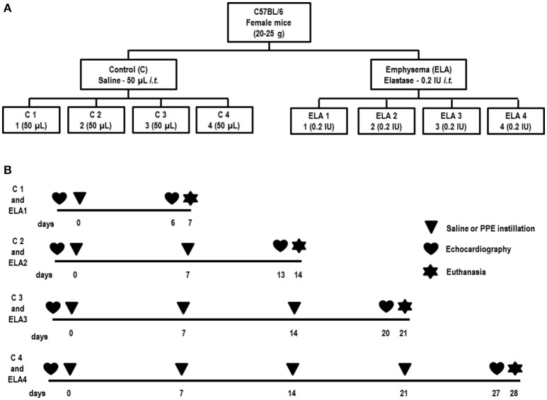
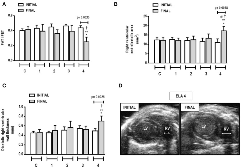
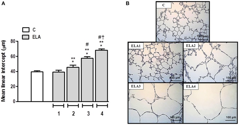
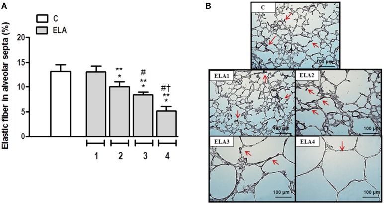
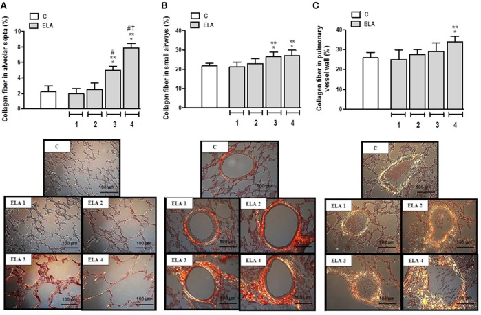
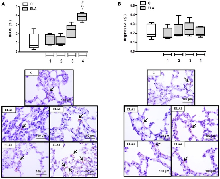
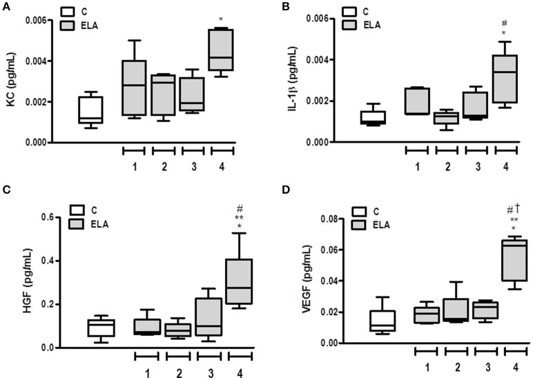
Many experimental models have been proposed to study the pathophysiological features of emphysema, as well as to search for new therapeutic approaches for acute or chronically injured lung parenchyma. We aimed to characterize an emphysema model induced by multiple instillations of elastase by tracking changes in inflammation, remodeling, and cardiac function after each instillation. Forty-eight C57BL/6 mice were randomly assigned across two groups. Emphysema (ELA) animals received 1, 2, 3, or 4 intratracheal instillations of pancreatic porcine elastase (PPE, 0.2 IU) with a 1-week interval between them. Controls (C) received saline following the same protocol. Before and after implementation of the protocol, animals underwent echocardiographic analysis. After the first instillation of PPE, the percentage of mononuclear cells in the lung parenchyma increased compared to C ( = 0.0001). The second instillation resulted in hyperinflated alveoli, increased mean linear intercept, and reduced elastic fiber content in lung parenchyma compared to C ( = 0.0197). Following the third instillation, neutrophils and collagen fiber content in alveolar septa and airways increased, whereas static lung elastance was reduced compared to C ( = 0.0094). After the fourth instillation, the percentage of M1 macrophages in lungs; levels of interleukin-1β (IL-1β), keratinocyte-derived chemokine, hepatocyte growth factor (HGF), and vascular endothelial growth factor (VEGF); and collagen fiber content in the pulmonary vessel wall were increased compared to C ( = 0.0096). At this time point, pulmonary arterial hypertension was apparent, with increased diastolic right ventricular wall thickness. In conclusion, the initial phase of emphysema was characterized by lung inflammation with predominance of mononuclear cells, whereas at the late stage, impairment of pulmonary and cardiovascular functions was observed. This model enables analysis of therapies at different time points during controlled progression of emphysema. Accordingly, early interventions could focus on the inflammatory process, while late interventions should focus on restoring cardiorespiratory function.
为了研究肺气肿的病理生理特征,并寻找针对急性或慢性损伤肺实质的新治疗方法,人们提出了许多实验模型。我们的目标是通过追踪每次滴注后炎症、重塑和心脏功能的变化,来表征多次滴注弹性蛋白酶诱导的肺气肿模型。48只C57BL/6小鼠被随机分为两组。肺气肿(ELA)组动物接受1、2、3或4次气管内滴注猪胰弹性蛋白酶(PPE,0.2 IU),每次滴注间隔1周。对照组(C)按照相同方案接受生理盐水滴注。在方案实施前后,对动物进行超声心动图分析。首次滴注PPE后,肺实质中单核细胞的百分比与C组相比增加(P = 0.0001)。第二次滴注导致肺泡过度充气,平均线性截距增加,与C组相比肺实质中弹性纤维含量减少(P = 0.0197)。第三次滴注后,肺泡间隔和气道中的中性粒细胞和胶原纤维含量增加,而与C组相比静态肺弹性降低(P = 0.0094)。第四次滴注后,与C组相比,肺中M1巨噬细胞的百分比、白细胞介素-1β(IL-1β)、角质形成细胞衍生趋化因子、肝细胞生长因子(HGF)和血管内皮生长因子(VEGF)的水平以及肺血管壁中的胶原纤维含量增加(P = 0.0096)。此时,肺动脉高压明显,舒张期右心室壁厚度增加。总之,肺气肿的初始阶段以单核细胞为主的肺部炎症为特征,而在晚期,则观察到肺和心血管功能受损。该模型能够在肺气肿的可控进展过程中分析不同时间点的治疗方法。因此,早期干预应侧重于炎症过程,而晚期干预应侧重于恢复心肺功能。