Mursch Kay, Scholz Martin, Brück Wolfgang, Behnke-Mursch Julianne
Department of Neurosurgery, Zentralklinik, Bad Berka, Germany.
Department of Neurosurgery, Klinikum Duisburg, Duisburg, Germany.
Ultrasonography. 2017 Jan;36(1):60-65. doi: 10.14366/usg.16015. Epub 2016 Aug 8.
The aim of this study was to investigate whether intraoperative ultrasonography (IOUS) helped the surgeon navigate towards the tumor as seen in preoperative magnetic resonance imaging and whether IOUS was able to distinguish between tumor margins and the surrounding tissue.
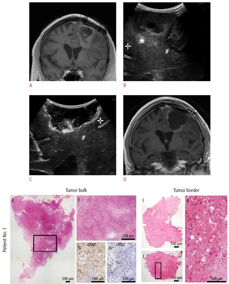
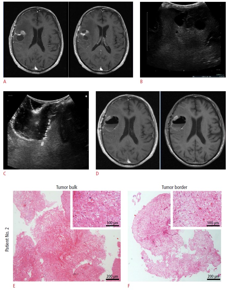
Twenty-five patients suffering from high-grade gliomas who were previously treated by surgery and radiotherapy were included. Intraoperatively, two histopathologic samples were obtained a sample of unequivocal tumor tissue (according to anatomical landmarks and the surgeon's visual and tactile impressions) and a small tissue sample obtained using a navigated needle when the surgeon decided to stop the resection. This specimen was considered to be a boundary specimen, where no tumor tissue was apparent. The decision to take the second sample was not influenced by IOUS. The effect of IOUS was analyzed semi-quantitatively.
All 25 samples of unequivocal tumor tissue were histopathologically classified as tumor tissue and were hyperechoic on IOUS. Of the boundary specimens, eight were hypoechoic. Only one harbored tumor tissue (P=0.150). Seventeen boundaries were moderately hyperechoic, and these samples contained all possible histological results (i.e., tumor, infiltration, or no tumor).
During surgery performed on relapsed, irradiated, high-grade gliomas, IOUS provided a reliable method of navigating towards the core of the tumor. At borders, it did not reliably distinguish between remnants or tumor-free tissue, but hypoechoic areas seldom contained tumor tissue.
本研究旨在调查术中超声检查(IOUS)是否有助于外科医生像在术前磁共振成像中看到的那样朝着肿瘤进行导航,以及IOUS是否能够区分肿瘤边缘与周围组织。
纳入25例先前接受过手术和放疗的高级别胶质瘤患者。术中获取两份组织病理学样本:一份明确为肿瘤组织的样本(根据解剖标志以及外科医生的视觉和触觉印象),另一份是当外科医生决定停止切除时使用导航针获取的小组织样本。该标本被视为边界标本,在其中未发现明显的肿瘤组织。获取第二份样本的决定不受IOUS影响。对IOUS的效果进行半定量分析。
所有25份明确为肿瘤组织的样本在组织病理学上均被分类为肿瘤组织,并且在IOUS上呈高回声。在边界标本中,8份为低回声。只有1份含有肿瘤组织(P = 0.150)。17份边界为中度高回声,这些样本包含了所有可能的组织学结果(即肿瘤、浸润或无肿瘤)。
在对复发、接受过放疗的高级别胶质瘤进行手术时,IOUS提供了一种可靠的朝着肿瘤核心进行导航的方法。在边界处,它不能可靠地区分残留组织或无肿瘤组织,但低回声区域很少含有肿瘤组织。