Division of Gastrointestinal Surgery, Nottingham Digestive Diseases Centre, Faculty of Medicine and Health Sciences, University of Nottingham, E Floor, West Block, Queen's Medical Centre, Derby Rd, Nottingham NG7 2UH, UK.
Research & Development Department, Lincoln Breast Unit, Lincoln County Hospital, Greetwell Road, Lincoln LN2 5QY, UK.
J Immunol Res. 2016;2016:4757405. doi: 10.1155/2016/4757405. Epub 2016 Sep 29.
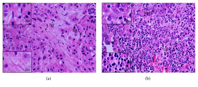
The tumour microenvironment consists of malignant cells, stroma, and immune cells. Prominent tumour-infiltrating lymphocytes (TILs) in breast cancer are associated with a good prognosis and are predictors of a pathological complete response (pCR) with neoadjuvant chemotherapy (NAC). The contribution of different T effector/regulatory cells and cytokines to tumour cell death with NAC requires further characterisation and was investigated in this study. Breast tumours from 33 women with large and locally advanced breast cancers undergoing NAC were immunohistochemically (intratumoural, stromal) assessed for T cell subsets and cytokine expression using labelled antibodies, employing established semiquantitative methods. Prominent levels of TILs and CD4, CD8, and CTLA-4 (stromal) T cells and CD8 : FOXP3 ratios were associated with a significant pCR; no association was seen with FOXP3, CTLA-4 (intratumoural), and PD-1 T cells. NAC significantly reduced CD4, FOXP3, CTLA-4 (stromal) (concurrently blood FOXP3, CTLA-4 Tregs), and PD-1 T cells; no reduction was seen with CD8 and CTLA-4 (intratumoural) T cells. High post-NAC tumour levels of FOXP3 T cells, IL-10, and IL-17 were associated with a failed pCR. Our study has characterised further the contribution of T effector/regulatory cells and cytokines to tumour cell death with NAC.
肿瘤微环境由恶性细胞、基质和免疫细胞组成。乳腺癌中浸润的肿瘤淋巴细胞(TILs)较多与预后良好相关,是新辅助化疗(NAC)后病理完全缓解(pCR)的预测因子。不同的 T 效应/调节细胞和细胞因子对 NAC 中肿瘤细胞死亡的贡献需要进一步阐明,本研究对此进行了探讨。对 33 例接受 NAC 的大且局部晚期乳腺癌女性的肿瘤组织进行免疫组化(肿瘤内、基质)评估,采用标记抗体,采用既定的半定量方法评估 T 细胞亚群和细胞因子表达。TILs 和 CD4、CD8 和 CTLA-4(基质)T 细胞以及 CD8:FOXP3 比值水平较高与显著的 pCR 相关;FOXP3、CTLA-4(肿瘤内)和 PD-1 T 细胞与 pCR 无相关性。NAC 显著降低了 CD4、FOXP3、CTLA-4(基质)(同时降低血液 FOXP3、CTLA-4 Tregs)和 PD-1 T 细胞;CD8 和 CTLA-4(肿瘤内)T 细胞没有减少。NAC 后肿瘤组织中 FOXP3 T 细胞、IL-10 和 IL-17 水平较高与 pCR 失败相关。本研究进一步阐明了 T 效应/调节细胞和细胞因子对 NAC 中肿瘤细胞死亡的贡献。