Syed Khaja Azharuddin Sajid, Toor Salman M, El Salhat Haytham, Faour Issam, Ul Haq Navid, Ali Bassam R, Elkord Eyad
Cancer Research Center, Qatar Biomedical Research Institute, College of Science and Engineering, Hamad Bin Khalifa University, Qatar Foundation, Doha, Qatar.
Department of Medical Microbiology and Immunology, College of Medicine and Health Sciences, United Arab Emirates University, Al Ain, United Arab Emirates.
Oncotarget. 2017 May 16;8(20):33159-33171. doi: 10.18632/oncotarget.16565.
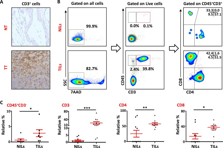
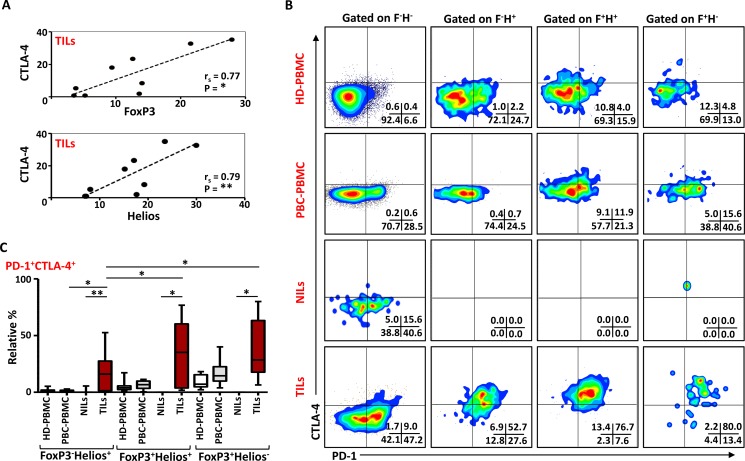
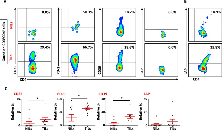
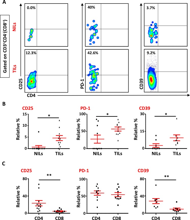
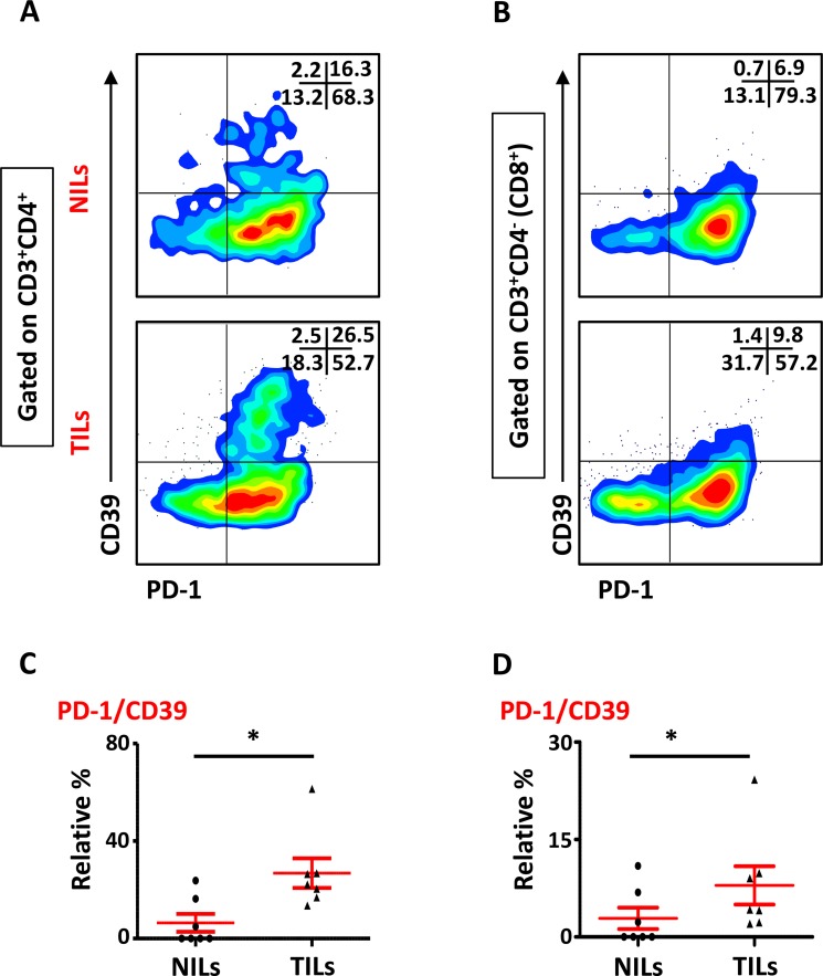
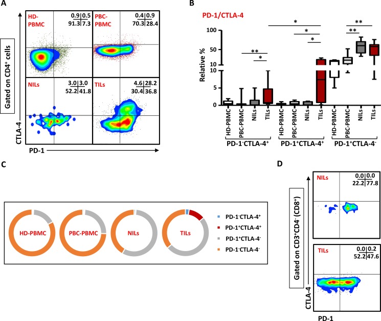
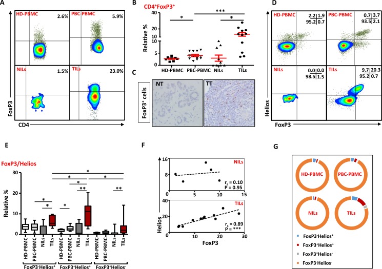
Immunosuppressive cells such as regulatory T cells (Tregs) have an ambiguous role in breast cancer prognosis, with studies reporting both positive and negative correlations between Treg infiltration and prognosis. This discrepancy could be due to the different immunosuppressive molecules present in these cells. In the present study, we phenotypically characterize different Treg subsets infiltrating the tumor microenvironment (TME), compared to adjacent normal tissue and peripheral blood of primary breast cancer (PBC) patients. We report that the majority of tumor-infiltrating CD4+ and CD8+ T cells have terminally exhaustive phenotype as assessed by CD39 and PD-1 expressions. We also show that Tregs are accumulated in breast TME compared to normal tissue. Further characterization of Tregs showed that these are mainly FoxP3+Helios+ and express high levels of CTLA-4 and PD-1. This preferential accumulation of FoxP3+Helios+ Treg subset with co-expression of different immune inhibitory molecules might have a negative effect on breast cancer prognosis. Taken together, our results suggest that breast tumor cells might utilize Tregs, and different suppressive pathways involving CD39, PD-1 and CTLA-4 molecules in creating an immune-subversive environment for them to survive, and a dual blockade of these immunosuppressive molecules might be considered as an effective method in breast cancer treatment.
免疫抑制细胞,如调节性T细胞(Tregs),在乳腺癌预后中具有模糊的作用,研究报告Treg浸润与预后之间存在正相关和负相关。这种差异可能是由于这些细胞中存在不同的免疫抑制分子。在本研究中,我们对浸润肿瘤微环境(TME)的不同Treg亚群进行了表型特征分析,并与原发性乳腺癌(PBC)患者的相邻正常组织和外周血进行了比较。我们报告说,通过CD39和PD-1表达评估,大多数肿瘤浸润性CD4+和CD8+ T细胞具有终末耗竭表型。我们还表明,与正常组织相比,Tregs在乳腺TME中积累。Tregs的进一步特征表明,这些主要是FoxP3+Helios+,并表达高水平的CTLA-4和PD-1。这种FoxP3+Helios+ Treg亚群的优先积累以及不同免疫抑制分子的共表达可能对乳腺癌预后产生负面影响。综上所述,我们的结果表明,乳腺肿瘤细胞可能利用Tregs以及涉及CD39、PD-1和CTLA-4分子的不同抑制途径来创造一个免疫颠覆环境以供它们生存,对这些免疫抑制分子的双重阻断可能被认为是乳腺癌治疗的有效方法。