Park Dae Woo, Sebastiani Andrea, Yap Choon Hwai, Simon Marc A, Kim Kang
Center for Ultrasound Molecular Imaging and Therapeutics, Department of Medicine, University of Pittsburgh School of Medicine, Pittsburgh, Pennsylvania, United States of America.
Heart and Vascular Institute, University of Pittsburgh Medical Center (UPMC), Pittsburgh, Pennsylvania, United States of America.
PLoS One. 2016 Oct 25;11(10):e0165320. doi: 10.1371/journal.pone.0165320. eCollection 2016.
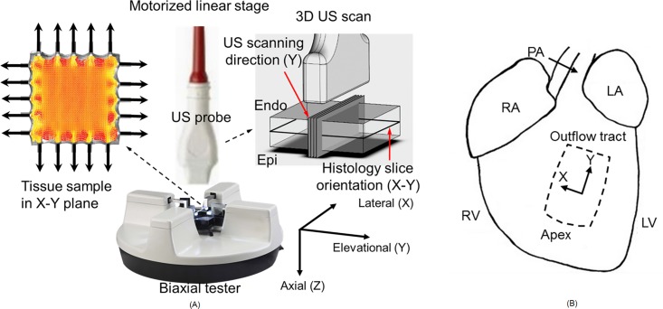
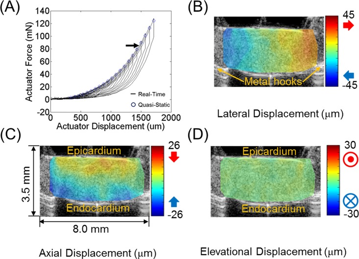
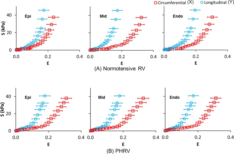
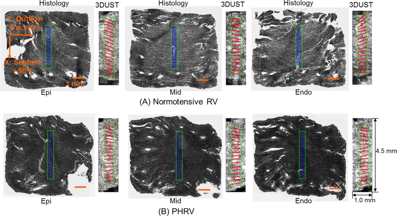
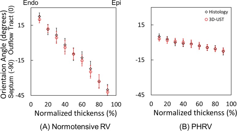
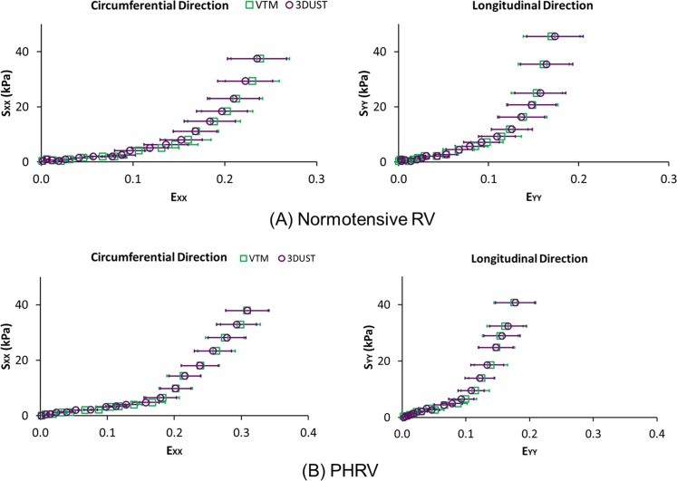
Mechanical and structural changes of right ventricular (RV) in response to pulmonary hypertension (PH) are inadequately understood. While current standard biaxial testing provides information on the mechanical behavior of RV tissues using surface markers, it is unable to fully assess structural and mechanical properties across the full tissue thickness. In this study, the mechanical and structural properties of normotensive and pulmonary hypertension right ventricular (PHRV) myocardium through its full thickness were examined using mechanical testing combined with 3D ultrasound speckle tracking (3D-UST). RV pressure overload was induced in Sprague-Dawley rats by pulmonary artery (PA) banding. The second Piola-Kirchhoff stress tensors and Green-Lagrangian strain tensors were computed in the RV myocardium using the biaxial testing combined with 3D-UST. A previously established non-linear curve-fitting algorithm was applied to fit experimental data to a Strain Energy Function (SEF) for computation of myofiber orientation. The fiber orientations obtained by the biaxial testing with 3D-UST compared well with the fiber orientations computed from the histology. In addition, the re-orientation of myofiber in the right ventricular free wall (RVFW) along longitudinal direction (apex-to-outflow-tract direction) was noticeable in response to PH. For normotensive RVFW samples, the average fiber orientation angles obtained by 3D-UST with biaxial test spiraled from 20° at the endo-cardium to -42° at the epi-cardium (Δ = 62°). For PHRV samples, the average fiber orientation angles obtained by 3D-UST with biaxial test had much less spiral across tissue thickness: 3° at endo-cardium to -7° at epi-cardium (Δ = 10°, P<0.005 compared to normotensive).
右心室(RV)对肺动脉高压(PH)的机械和结构变化尚未得到充分了解。虽然目前的标准双轴测试使用表面标记提供了有关RV组织机械行为的信息,但它无法全面评估整个组织厚度的结构和机械性能。在本研究中,通过机械测试结合三维超声斑点追踪(3D-UST),对正常血压和肺动脉高压右心室(PHRV)心肌全层的机械和结构特性进行了研究。通过肺动脉(PA)环扎在Sprague-Dawley大鼠中诱导RV压力过载。使用双轴测试结合3D-UST计算RV心肌中的第二Piola-Kirchhoff应力张量和Green-Lagrangian应变张量。应用先前建立 的非线性曲线拟合算法将实验数据拟合到应变能函数(SEF),以计算肌纤维方向。通过双轴测试与3D-UST获得的纤维方向与从组织学计算得到的纤维方向比较良好。此外,响应PH,右心室游离壁(RVFW)中的肌纤维沿纵向(心尖到流出道方向)重新定向很明显。对于正常血压的RVFW样本,通过3D-UST与双轴测试获得的平均纤维方向角从心内膜处的20°螺旋到心外膜处的-42°(Δ=62°)。对于PHRV样本,通过3D-UST与双轴测试获得的平均纤维方向角在整个组织厚度上的螺旋程度要小得多:心内膜处为3°,心外膜处为-7°(Δ=10°,与正常血压相比P<0.005)。