• 文献检索
  • 文档翻译
  • 深度研究
  • 学术资讯
  • Suppr Zotero 插件Zotero 插件
  • 邀请有礼
  • 套餐&价格
  • 历史记录
应用&插件
Suppr Zotero 插件Zotero 插件浏览器插件Mac 客户端Windows 客户端微信小程序
定价
高级版会员购买积分包购买API积分包
服务
文献检索文档翻译深度研究API 文档MCP 服务
关于我们
关于 Suppr公司介绍联系我们用户协议隐私条款
关注我们

Suppr 超能文献

核心技术专利:CN118964589B侵权必究
粤ICP备2023148730 号-1Suppr @ 2026

文献检索

告别复杂PubMed语法,用中文像聊天一样搜索,搜遍4000万医学文献。AI智能推荐,让科研检索更轻松。

立即免费搜索

文件翻译

保留排版,准确专业,支持PDF/Word/PPT等文件格式,支持 12+语言互译。

免费翻译文档

深度研究

AI帮你快速写综述,25分钟生成高质量综述,智能提取关键信息,辅助科研写作。

立即免费体验

使用锥形束计算机断层扫描对伊朗人群中上颌第一磨牙的解剖结构和根管形态进行年龄和性别的调查。

Survey of Anatomy and Root Canal Morphology of Maxillary First Molars Regarding Age and Gender in an Iranian Population Using Cone-Beam Computed Tomography.

作者信息

Naseri Mandana, Safi Yaser, Akbarzadeh Baghban Alireza, Khayat Akbar, Eftekhar Leila

机构信息

Department of Endodontics, Dental School, Shahid Beheshti University of Medical Sciences, Tehran, Iran.

Oral and Maxillofacial Radiology Department, Dental School, Shahid Beheshti University of Medical Sciences, Tehran, Iran.

出版信息

Iran Endod J. 2016 Fall;11(4):298-303. doi: 10.22037/iej.2016.8.

DOI:10.22037/iej.2016.8
PMID:27790259
原文链接:https://pmc.ncbi.nlm.nih.gov/articles/PMC5069906/
Abstract

INTRODUCTION

The purpose of this study was to investigate the root and canal morphology of maxillary first molars with regards to patients' age and gender with cone-beam computed tomography (CBCT).

METHODS AND MATERIALS

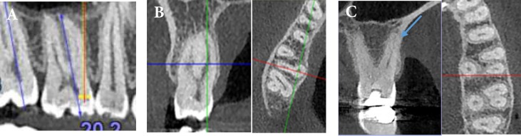
A total of 149 CBCT scans from 92 (67.1%) female and 57 (31.3%) male patients with mean age of 40.5 years were evaluated. Tooth length, presence of root fusion, number of the roots and canals, canal types based on Vertucci's classification, deviation of root and apical foramen in coronal and sagittal planes and the correlation of all items with gender and age were recorded. The Mann Whitney U, Kruskal Wallis and Fisher's exact tests were used to analyze these items.

RESULTS

The rate of root fusion was 1.3%. Multiple canals were present in the following frequencies: four canals 78.5%, five canals 11.4% and three canals 10.1%. Additional canal was detected in 86.6% of mesiobuccal roots in which Vertucci's type configuration was the most prevalent followed by type and . Type was the most common one in distobuccal and palatal roots. There was no statistically significant difference in the canal configurations in relation to gender and age as well as the incidence root or canal numbers (>0.05). The mean tooth length was 19.3 and 20.3 mm in female and male patients, respectively which was statistically significant (<0.05). Evaluation of root deviation showed that most commonly, a general pattern of straight-distal in the mesiobuccal and straight-straight for distobuccal and palatal roots occurred. In mesiobuccal roots, straight and distal deviations were more dominant in male and female, respectively (<0.05). The prevalence of apical foramen deviation in mesiobuccal and palatal roots statistically differed with gender.

CONCLUSION

The root and canal configuration of Iranian population showed different features from those of other populations.

摘要

引言

本研究的目的是利用锥形束计算机断层扫描(CBCT),研究上颌第一磨牙的牙根和根管形态与患者年龄及性别的关系。

方法和材料

共评估了149例CBCT扫描,这些扫描来自92例(67.1%)女性和57例(31.3%)男性患者,平均年龄为40.5岁。记录牙齿长度、牙根融合情况、牙根和根管数量、基于维尔图奇分类的根管类型、牙根和根尖孔在冠状面和矢状面的偏差以及所有项目与性别和年龄的相关性。采用曼-惠特尼U检验、克鲁斯卡尔-沃利斯检验和费舍尔精确检验来分析这些项目。

结果

牙根融合率为1.3%。多根管出现的频率如下:四根管78.5%,五根管11.4%,三根管10.1%。在86.6%的近中颊根中检测到额外根管,其中维尔图奇分类的构型最为常见,其次是构型和构型。构型是远中颊根和腭根中最常见的类型。根管构型在性别和年龄方面以及牙根或根管数量的发生率上无统计学显著差异(>0.05)。女性和男性患者的平均牙齿长度分别为19.3毫米和20.3毫米,具有统计学显著性差异(<0.05)。牙根偏差评估显示,最常见的情况是,近中颊根呈直-远中方向,远中颊根和腭根呈直-直方向。在近中颊根中,直向和远中向偏差在男性和女性中分别更为明显(<0.05)。近中颊根和腭根根尖孔偏差的发生率在性别上有统计学差异。

结论

伊朗人群的牙根和根管形态与其他人群表现出不同特征。

https://cdn.ncbi.nlm.nih.gov/pmc/blobs/a266/5069906/93b6c1b7614f/iej-11-298-g001.jpg

相似文献

1
Survey of Anatomy and Root Canal Morphology of Maxillary First Molars Regarding Age and Gender in an Iranian Population Using Cone-Beam Computed Tomography.使用锥形束计算机断层扫描对伊朗人群中上颌第一磨牙的解剖结构和根管形态进行年龄和性别的调查。
Iran Endod J. 2016 Fall;11(4):298-303. doi: 10.22037/iej.2016.8.
2
Root Canal Morphology of Maxillary Second Molars according to Age and Gender in a Selected Iranian Population: A Cone-Beam Computed Tomography Evaluation.伊朗特定人群中上颌第二磨牙根管形态与年龄和性别的关系:锥形束计算机断层扫描评估
Iran Endod J. 2018 Summer;13(3):373-380. doi: 10.22037/iej.v13i3.19278.
3
Evaluation of root canal morphology of maxillary molars using cone beam computed tomography.使用锥形束计算机断层扫描评估上颌磨牙的根管形态
Pak J Med Sci. 2015 Mar-Apr;31(2):426-30. doi: 10.12669/pjms.312.6753.
4
Root Morphology of the Maxillary First and Second Molars in an Iranian Population Using Cone Beam Computed Tomography.使用锥形束计算机断层扫描技术对上颌第一和第二磨牙在伊朗人群中的牙根形态进行研究。
J Dent (Tehran). 2017 May;14(3):115-122.
5
Cone-beam computed tomography evaluation of maxillary first and second molars in Iranian population: a morphological study.伊朗人群上颌第一和第二磨牙的锥形束计算机断层扫描评估:一项形态学研究。
Iran Endod J. 2014 Summer;9(3):190-4. Epub 2014 Jul 5.
6
A Cone-beam Computed Tomographic Study of Root and Canal Morphology of Maxillary First and Second Permanent Molars in a Thai Population.一项泰国人群上颌第一和第二恒磨牙根管及形态的锥形束 CT 研究。
J Endod. 2018 Jan;44(1):56-61. doi: 10.1016/j.joen.2017.08.020. Epub 2017 Oct 20.
7
Evaluation of the relationship between buccolingual width of mesiobuccal root and root canal morphology of maxillary first molars by cone-beam computed tomography.锥形束计算机断层扫描评估上颌第一磨牙近中颊根颊舌径与根管形态的关系
Dent Res J (Isfahan). 2022 Jan 28;19:5. doi: 10.4103/1735-3327.336690. eCollection 2022.
8
Evaluation of root canal configuration of maxillary molars in a Brazilian population using cone-beam computed tomographic imaging: an in vivo study.使用锥形束计算机断层扫描成像评估巴西人群上颌磨牙的根管形态:一项体内研究。
J Endod. 2014 Feb;40(2):173-6. doi: 10.1016/j.joen.2013.10.002. Epub 2013 Nov 9.
9
Root and Canal Morphology of Maxillary Teeth in an Iranian Subpopulation Residing in Western Iran Using Cone-Beam Computed Tomography.使用锥形束计算机断层扫描对居住在伊朗西部的一个伊朗亚人群上颌牙的根管形态进行研究。
Iran Endod J. 2020 Winter;15(1):31-37. doi: 10.22037/iej.v15i1.25386.
10
Root canal morphology of permanent teeth in a Malaysian subpopulation using cone-beam computed tomography.马来西亚人群恒牙根尖形态的锥形束 CT 研究。
BMC Oral Health. 2019 Jan 14;19(1):14. doi: 10.1186/s12903-019-0710-z.

引用本文的文献

1
Prevalence of second mesiobuccal canal in maxillary molars of Iranian population: A systematic review with meta-analysis.伊朗人群上颌磨牙近中颊侧第二根管的患病率:一项Meta分析的系统评价
PLoS One. 2025 Jul 11;20(7):e0327006. doi: 10.1371/journal.pone.0327006. eCollection 2025.
2
Prevalence of Untreated Canals and Their Association with Periapical Periodontitis Using Cone-beam Computed Tomography.使用锥形束计算机断层扫描技术评估未经治疗根管的患病率及其与根尖周炎的关联。
Iran Endod J. 2024;19(3):150-157. doi: 10.22037/iej.v19i3.42440.
3
CBCT Evaluation of Root Canal Morphology of Maxillary First Premolar in Saudi Subpopulation.

本文引用的文献

1
Root Canal Morphology of Permanent Mandibular Premolars in Iranian Population: A Systematic Review.伊朗人群恒牙下颌前磨牙的根管形态:一项系统评价
Iran Endod J. 2016 Summer;11(3):150-6. doi: 10.7508/iej.2016.03.001. Epub 2016 May 1.
2
The applications of cone-beam computed tomography in endodontics: a review of literature.锥形束计算机断层扫描在牙髓病学中的应用:文献综述
Iran Endod J. 2015 Winter;10(1):16-25. Epub 2014 Dec 24.
3
Endodontic treatment of a double-rooted maxillary second molar with four canals: a case report.
沙特亚人群上颌第一前磨牙根管形态的锥形束计算机断层扫描评估
J Pharm Bioallied Sci. 2024 Apr;16(Suppl 2):S1619-S1622. doi: 10.4103/jpbs.jpbs_1048_23. Epub 2024 Apr 16.
4
Retrospective study of the morphology of third maxillary molars among the population of Lower Silesia based on analysis of cone beam computed tomography.基于锥形束计算机断层扫描分析的下西里西亚人群第三磨牙形态的回顾性研究。
PLoS One. 2024 Feb 23;19(2):e0299123. doi: 10.1371/journal.pone.0299123. eCollection 2024.
5
Cone-Beam Computed Tomography Assessment of Prevalence of Procedural Errors in Maxillary Posterior Teeth.锥形束计算机断层扫描评估上颌后牙操作失误的发生率。
Biomed Res Int. 2023 Dec 12;2023:4439890. doi: 10.1155/2023/4439890. eCollection 2023.
6
Variables That Affect the Ability to Find the Second Mesiobuccal Root Canals in Maxillary Molars.影响上颌磨牙近中颊根第二根管发现能力的因素
Iran Endod J. 2023;18(4):248-253. doi: 10.22037/iej.v18i4.42260.
7
Root Canal Treatment of a Geminated Maxillary Second Molar with C-shaped Canal System: A Case Report.伴有C形根管系统的上颌第二磨牙融合根根管治疗:一例报告
Iran Endod J. 2023;18(3):181-185. doi: 10.22037/iej.v18i3.41384.
8
A Scoping Review of Highly Cited Published Articles in the Iranian Endodontic Journal: A Reflection of Trends in Endodontic Research.对《伊朗牙髓病学杂志》高被引发表文章的范围综述:牙髓病学研究趋势的反映
Iran Endod J. 2023;18(3):126-133. doi: 10.22037/iej.v18i2.42524.
9
A CBCT Study to Evaluate the Root and Canal Morphology of Permanent Maxillary First Molars in Children.一项评估儿童恒牙上颌第一磨牙牙根和根管形态的锥形束计算机断层扫描(CBCT)研究。
Int J Clin Pediatr Dent. 2022 Sep-Oct;15(5):509-513. doi: 10.5005/jp-journals-10005-2441.
10
The morphology of permanent maxillary first molars evaluated by cone-beam computed tomography among a Yemeni population.利用锥形束计算机断层扫描评估也门人群中恒上颌第一磨牙的形态。
BMC Oral Health. 2023 Jan 27;23(1):46. doi: 10.1186/s12903-023-02752-2.
Iran Endod J. 2014 Fall;9(4):304-6. Epub 2014 Oct 7.
4
Cone-beam computed tomography evaluation of maxillary first and second molars in Iranian population: a morphological study.伊朗人群上颌第一和第二磨牙的锥形束计算机断层扫描评估:一项形态学研究。
Iran Endod J. 2014 Summer;9(3):190-4. Epub 2014 Jul 5.
5
Evaluation of root and canal morphology of maxillary permanent first molars in a North American population by cone-beam computed tomography.通过锥形束计算机断层扫描评估北美人群上颌第一恒磨牙的根管形态。
J Endod. 2014 May;40(5):635-9. doi: 10.1016/j.joen.2014.02.002. Epub 2014 Mar 29.
6
Two symmetrical maxillary first molars with two disto-buccal root canals.两颗对称的上颌第一磨牙,各有两条远中颊根根管。
Iran Endod J. 2006 Summer;1(2):73-7. Epub 2006 Jul 1.
7
Evaluation of root canal configuration of maxillary molars in a Brazilian population using cone-beam computed tomographic imaging: an in vivo study.使用锥形束计算机断层扫描成像评估巴西人群上颌磨牙的根管形态:一项体内研究。
J Endod. 2014 Feb;40(2):173-6. doi: 10.1016/j.joen.2013.10.002. Epub 2013 Nov 9.
8
An ex vivo comparison of digital radiography and cone-beam and micro computed tomography in the detection of the number of canals in the mesiobuccal roots of maxillary molars.上颌磨牙近颊根根管数的数字射线摄影与锥形束和微计算机断层扫描的体外比较。
J Endod. 2013 Jul;39(7):901-5. doi: 10.1016/j.joen.2013.01.010. Epub 2013 May 14.
9
Second canal in mesiobuccal root of maxillary molars is correlated with root third and patient age: a cone-beam computed tomographic study.上颌磨牙近颊根第二根管与根管第三分支和患者年龄相关:锥形束 CT 研究。
J Endod. 2013 May;39(5):588-92. doi: 10.1016/j.joen.2013.01.003. Epub 2013 Mar 7.
10
Morphology of maxillary first and second molars analyzed by cone-beam computed tomography in a korean population: variations in the number of roots and canals and the incidence of fusion.锥形束计算机断层扫描分析韩国人群上颌第一、二磨牙的形态:牙根和根管数量的变异及融合的发生率。
J Endod. 2012 Aug;38(8):1063-8. doi: 10.1016/j.joen.2012.04.025. Epub 2012 Jun 20.