• 文献检索
  • 文档翻译
  • 深度研究
  • 学术资讯
  • Suppr Zotero 插件Zotero 插件
  • 邀请有礼
  • 套餐&价格
  • 历史记录
应用&插件
Suppr Zotero 插件Zotero 插件浏览器插件Mac 客户端Windows 客户端微信小程序
定价
高级版会员购买积分包购买API积分包
服务
文献检索文档翻译深度研究API 文档MCP 服务
关于我们
关于 Suppr公司介绍联系我们用户协议隐私条款
关注我们

Suppr 超能文献

核心技术专利:CN118964589B侵权必究
粤ICP备2023148730 号-1Suppr @ 2026

文献检索

告别复杂PubMed语法,用中文像聊天一样搜索,搜遍4000万医学文献。AI智能推荐,让科研检索更轻松。

立即免费搜索

文件翻译

保留排版,准确专业,支持PDF/Word/PPT等文件格式,支持 12+语言互译。

免费翻译文档

深度研究

AI帮你快速写综述,25分钟生成高质量综述,智能提取关键信息,辅助科研写作。

立即免费体验

使用锥形束计算机断层扫描技术对上颌第一和第二磨牙在伊朗人群中的牙根形态进行研究。

Root Morphology of the Maxillary First and Second Molars in an Iranian Population Using Cone Beam Computed Tomography.

作者信息

Ghoncheh Zahra, Zade Behrang Moghaddam, Kharazifard Mohammad Javad

机构信息

Assistant Professor, Department of Oral and Maxillofacial Radiology, School of Dentistry, Tehran University of Medical Sciences, International Campus, Tehran, Iran.

Adjunct Professor, Department of Oral and Maxillofacial Radiology, School of Dentistry, Tehran University of Medical Sciences, International Campus, Tehran, Iran.

出版信息

J Dent (Tehran). 2017 May;14(3):115-122.

PMID:29167682
原文链接:https://pmc.ncbi.nlm.nih.gov/articles/PMC5694843/
Abstract

OBJECTIVES

This study sought to assess the root morphology and root canal anatomy of the maxillary first and second molars in an Iranian population using cone-beam computed tomography (CBCT).

MATERIALS AND METHODS

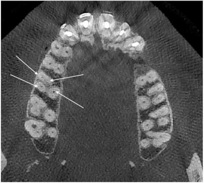
Sound fully-developed maxillary first (n=345) and second (n=423) molars were evaluated on 450 CBCT scans ordered for pre-operative assessment for implant placement. The (I) number of roots and their morphology (II) number of canals per root, (III) canal configuration and presence of a second mesiobuccal canal according to the Vertucci's classification and (IV) unilateral or bilateral occurrence of a second mesiobuccal canal (MB2) were evaluated.

RESULTS

Single roots were found in 1.1% of the first and 11.3% of the second molars. Four separate roots were identified in 0.5% of the first molars; none of the second molars had four separate roots. First and second molars showed a higher prevalence of three separate roots of mesiobuccal, distobuccal and palatal with one canal in each root (54% and 86 %, respectively). The most common anatomical variation in the maxillary first molars was related to the configuration of the MB root; the root canal system of the maxillary second molars showed more anatomical variations.

CONCLUSIONS

Mesiobuccal roots of the maxillary molars had more variations in their canal system than the distobuccal or palatal roots. The root canal configuration of the maxillary second molars was more diverse than that of first molars; CBCT enhances mapping of the mesiobuccal root canal system with the potential to improve the quality of root canal treatment.

摘要

目的

本研究旨在使用锥形束计算机断层扫描(CBCT)评估伊朗人群上颌第一和第二磨牙的牙根形态及根管解剖结构。

材料与方法

对450例为种植术前评估而进行的CBCT扫描中的完好、发育完全的上颌第一磨牙(n = 345)和第二磨牙(n = 423)进行评估。评估内容包括:(I)牙根数量及其形态;(II)每根根管的数量;(III)根据韦尔图奇分类法的根管形态及第二近中颊根管的存在情况;(IV)第二近中颊根管(MB2)的单侧或双侧出现情况。

结果

在1.1%的第一磨牙和11.3%的第二磨牙中发现单根。在0.5%的第一磨牙中发现有四根独立的牙根;第二磨牙均无四根独立的牙根。第一和第二磨牙中,近中颊根、远中颊根和腭根三根独立且每根有一个根管的情况更为常见(分别为54%和86%)。上颌第一磨牙最常见的解剖变异与近中颊根的形态有关;上颌第二磨牙的根管系统显示出更多的解剖变异。

结论

上颌磨牙的近中颊根根管系统比远中颊根或腭根有更多变异。上颌第二磨牙的根管形态比第一磨牙更多样化;CBCT增强了近中颊根管系统的成像,有可能提高根管治疗的质量。

https://cdn.ncbi.nlm.nih.gov/pmc/blobs/e957/5694843/e0197fa0ae3b/JOD-14-115-g001.jpg

相似文献

1
Root Morphology of the Maxillary First and Second Molars in an Iranian Population Using Cone Beam Computed Tomography.使用锥形束计算机断层扫描技术对上颌第一和第二磨牙在伊朗人群中的牙根形态进行研究。
J Dent (Tehran). 2017 May;14(3):115-122.
2
Use of CBCT to identify the morphology of maxillary permanent molar teeth in a Chinese subpopulation.应用 CBCT 分析中国人群上颌恒磨牙的解剖形态。
Int Endod J. 2011 Feb;44(2):162-9. doi: 10.1111/j.1365-2591.2010.01826.x. Epub 2010 Nov 22.
3
A Cone-beam Computed Tomographic Study of Root and Canal Morphology of Maxillary First and Second Permanent Molars in a Thai Population.一项泰国人群上颌第一和第二恒磨牙根管及形态的锥形束 CT 研究。
J Endod. 2018 Jan;44(1):56-61. doi: 10.1016/j.joen.2017.08.020. Epub 2017 Oct 20.
4
Cone-beam computed tomography evaluation of maxillary first and second molars in Iranian population: a morphological study.伊朗人群上颌第一和第二磨牙的锥形束计算机断层扫描评估:一项形态学研究。
Iran Endod J. 2014 Summer;9(3):190-4. Epub 2014 Jul 5.
5
Evaluation of root canal configuration of maxillary molars in a Brazilian population using cone-beam computed tomographic imaging: an in vivo study.使用锥形束计算机断层扫描成像评估巴西人群上颌磨牙的根管形态:一项体内研究。
J Endod. 2014 Feb;40(2):173-6. doi: 10.1016/j.joen.2013.10.002. Epub 2013 Nov 9.
6
Evaluation of root canal morphology of maxillary molars using cone beam computed tomography.使用锥形束计算机断层扫描评估上颌磨牙的根管形态
Pak J Med Sci. 2015 Mar-Apr;31(2):426-30. doi: 10.12669/pjms.312.6753.
7
Evaluation of the relationship between buccolingual width of mesiobuccal root and root canal morphology of maxillary first molars by cone-beam computed tomography.锥形束计算机断层扫描评估上颌第一磨牙近中颊根颊舌径与根管形态的关系
Dent Res J (Isfahan). 2022 Jan 28;19:5. doi: 10.4103/1735-3327.336690. eCollection 2022.
8
Assessment of the presence of a second mesiobuccal canal in maxillary first molars according to the location of the main mesiobuccal canal-a micro-computed tomographic study.上颌第一磨牙第二近中颊根管存在情况的评估:基于主近中颊根管位置的分析-一项显微 CT 研究。
Clin Oral Investig. 2021 Jun;25(6):3937-3944. doi: 10.1007/s00784-020-03723-5. Epub 2021 Jan 6.
9
Analysis of the Root and Canal Morphologies in Maxillary First and Second Molars in a Chinese Population Using Cone-beam Computed Tomography.使用锥形束计算机断层扫描分析中国人群上颌第一和第二磨牙的根管形态
J Endod. 2016 May;42(5):696-701. doi: 10.1016/j.joen.2016.01.017. Epub 2016 Mar 16.
10
Root Canal Morphology of Maxillary Second Molars according to Age and Gender in a Selected Iranian Population: A Cone-Beam Computed Tomography Evaluation.伊朗特定人群中上颌第二磨牙根管形态与年龄和性别的关系:锥形束计算机断层扫描评估
Iran Endod J. 2018 Summer;13(3):373-380. doi: 10.22037/iej.v13i3.19278.

引用本文的文献

1
Prevalence of second mesiobuccal canal in maxillary molars of Iranian population: A systematic review with meta-analysis.伊朗人群上颌磨牙近中颊侧第二根管的患病率:一项Meta分析的系统评价
PLoS One. 2025 Jul 11;20(7):e0327006. doi: 10.1371/journal.pone.0327006. eCollection 2025.
2
Atypical Root Canal System Anatomy in a Permanent Upper First Molar: A Case Report.一例恒牙上颌第一磨牙的非典型根管系统解剖:病例报告
Cureus. 2025 Mar 18;17(3):e80760. doi: 10.7759/cureus.80760. eCollection 2025 Mar.
3
Root Morphology of First Permanent Molars Extracted from Adult Dental Patients of Similar Ethnicity in Dar-Es-Salaam, Tanzania.

本文引用的文献

1
Root canal configuration of permanent maxillary first molar in Khasi population of Meghalaya: An in vitro study.梅加拉亚邦卡西族人群上颌第一恒磨牙的根管形态:一项体外研究。
J Conserv Dent. 2014 Jul;17(4):359-63. doi: 10.4103/0972-0707.136511.
2
Cone-beam computed tomography evaluation of maxillary first and second molars in Iranian population: a morphological study.伊朗人群上颌第一和第二磨牙的锥形束计算机断层扫描评估:一项形态学研究。
Iran Endod J. 2014 Summer;9(3):190-4. Epub 2014 Jul 5.
3
Evaluation of root and canal morphology of maxillary permanent first molars in a North American population by cone-beam computed tomography.
坦桑尼亚达累斯萨拉姆地区成年同种族牙科患者拔除的第一恒磨牙的牙根形态
Int J Dent. 2025 Feb 28;2025:2531403. doi: 10.1155/ijod/2531403. eCollection 2025.
4
Anatomical Factors of the Anterior and Posterior Maxilla Affecting Immediate Implant Placement Based on Cone Beam Computed Tomography Analysis: A Narrative Review.基于锥形束计算机断层扫描分析的上颌前牙区和后牙区影响即刻种植的解剖学因素:一项叙述性综述
Diagnostics (Basel). 2024 Aug 5;14(15):1697. doi: 10.3390/diagnostics14151697.
5
Variables That Affect the Ability to Find the Second Mesiobuccal Root Canals in Maxillary Molars.影响上颌磨牙近中颊根第二根管发现能力的因素
Iran Endod J. 2023;18(4):248-253. doi: 10.22037/iej.v18i4.42260.
6
Evaluation of roots, root canal morphology, and bilateral symmetry of maxillary first molars in a Syrian subpopulation using cone beam computed tomography.应用锥形束 CT 评估叙利亚人群上颌第一磨牙的牙根、根管形态和双侧对称性。
Clin Exp Dent Res. 2023 Dec;9(6):1149-1155. doi: 10.1002/cre2.782. Epub 2023 Sep 11.
7
Endodontic Surgery of the Palatal Root of a Maxillary Molar Associated with Simultaneous Management of a Maxillary Sinus Lesion.上颌磨牙腭根牙髓外科手术联合上颌窦病变同期处理
Case Rep Dent. 2023 Jul 12;2023:9180800. doi: 10.1155/2023/9180800. eCollection 2023.
8
Non-surgical Root Canal Treatment of an Upper First Molar With an Unusual Morphology: A Case Report.上颌第一磨牙罕见形态的非手术根管治疗:病例报告
Cureus. 2023 May 30;15(5):e39725. doi: 10.7759/cureus.39725. eCollection 2023 May.
9
Automatic Classification System for Periapical Lesions in Cone-Beam Computed Tomography.根尖病变的锥形束计算机断层扫描自动分类系统。
Sensors (Basel). 2022 Aug 28;22(17):6481. doi: 10.3390/s22176481.
10
Evaluation of Root Anatomy and Canal Configuration of Human Permanent Maxillary First Molar Using Cone-Beam Computed Tomography: A Systematic Review.利用锥形束 CT 评估人上颌第一磨牙的牙根解剖和根管形态:系统评价。
Int J Environ Res Public Health. 2022 Aug 16;19(16):10160. doi: 10.3390/ijerph191610160.
通过锥形束计算机断层扫描评估北美人群上颌第一恒磨牙的根管形态。
J Endod. 2014 May;40(5):635-9. doi: 10.1016/j.joen.2014.02.002. Epub 2014 Mar 29.
4
Evaluation of root canal configuration of maxillary molars in a Brazilian population using cone-beam computed tomographic imaging: an in vivo study.使用锥形束计算机断层扫描成像评估巴西人群上颌磨牙的根管形态:一项体内研究。
J Endod. 2014 Feb;40(2):173-6. doi: 10.1016/j.joen.2013.10.002. Epub 2013 Nov 9.
5
Cone-beam computed tomography study of root and canal morphology of mandibular premolars in a western Chinese population.中国西部人群下颌前磨牙根管及形态的锥形束 CT 研究。
BMC Med Imaging. 2012 Jul 20;12:18. doi: 10.1186/1471-2342-12-18.
6
Morphology of maxillary first and second molars analyzed by cone-beam computed tomography in a korean population: variations in the number of roots and canals and the incidence of fusion.锥形束计算机断层扫描分析韩国人群上颌第一、二磨牙的形态:牙根和根管数量的变异及融合的发生率。
J Endod. 2012 Aug;38(8):1063-8. doi: 10.1016/j.joen.2012.04.025. Epub 2012 Jun 20.
7
Use of CBCT to identify the morphology of maxillary permanent molar teeth in a Chinese subpopulation.应用 CBCT 分析中国人群上颌恒磨牙的解剖形态。
Int Endod J. 2011 Feb;44(2):162-9. doi: 10.1111/j.1365-2591.2010.01826.x. Epub 2010 Nov 22.
8
Cone-beam computed tomography study of root and canal morphology of maxillary first and second molars in an Indian population.上颌第一、二磨牙根管及形态的锥形束 CT 研究。
J Endod. 2010 Oct;36(10):1622-7. doi: 10.1016/j.joen.2010.07.006. Epub 2010 Aug 30.
9
A cone-beam computed tomography study of maxillary first permanent molar root and canal morphology in a Chinese population.中国人上颌第一恒磨牙根管及形态的锥形束 CT 研究。
J Endod. 2010 Sep;36(9):1480-4. doi: 10.1016/j.joen.2010.06.018.
10
Dimension, anatomy and morphology of the mesiobuccal root canal system in maxillary molars.上颌磨牙近颊根管系统的维度、解剖和形态。
J Endod. 2010 Jun;36(6):985-9. doi: 10.1016/j.joen.2010.02.017. Epub 2010 Mar 19.