Petritsch B, Köstler H, Weng A M, Horn M, Gassenmaier T, Kunz A S, Weidemann F, Wanner C, Bley T A, Beer M
Department of Diagnostic and Interventional Radiology, University Hospital Würzburg, Oberdürrbacher Straße 6, 97080, Würzburg, Germany.
University of Würzburg, Comprehensive Heart Failure Center, 97080, Würzburg, Germany.
BMC Cardiovasc Disord. 2016 Oct 28;16(1):205. doi: 10.1186/s12872-016-0382-4.
Fabry disease is characterized by a progressive deposition of sphingolipids in different organ systems, whereby cardiac involvement leads to death. We hypothesize that lysosomal storage of sphingolipids in the heart as occurring in Fabry disease does not reflect in higher cardiac lipid concentrations detectable by H magnetic resonance spectroscopy (MRS) at 3 Tesla.
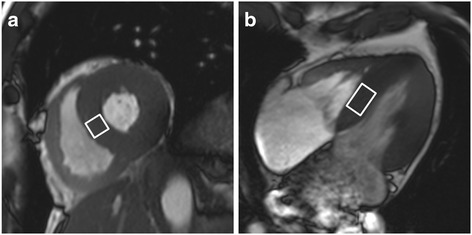
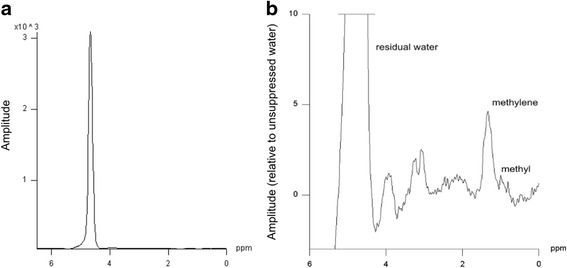
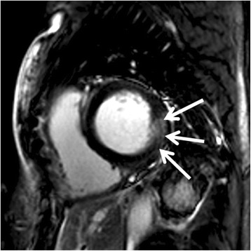
Myocardial lipid content was quantified in vivo by H-MRS in 30 patients (12 male, 18 female; 18 patients treated with enzyme replacement therapy) with genetically proven Fabry disease and in 30 healthy controls. The study protocol combined H-MRS with cardiac cine imaging and LGE MRI in a single examination.
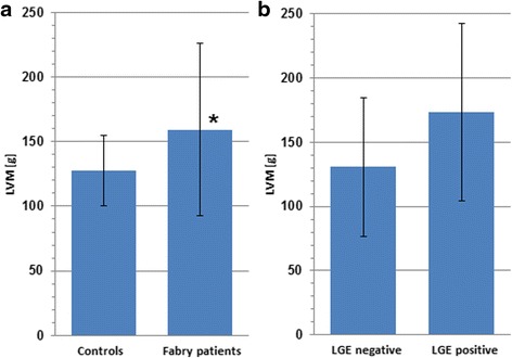
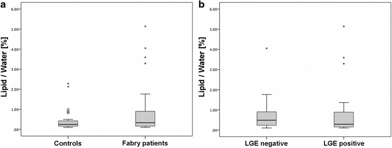
Myocardial lipid content was not significantly elevated in Fabry disease (p = 0.225). Left ventricular (LV) mass was significantly higher in patients suffering from Fabry disease compared to controls (p = 0.019). Comparison of patients without signs of myocardial fibrosis in MRI (LGE negative; n = 12) to patients with signs of fibrosis (LGE positive; n = 18) revealed similar myocardial lipid content in both groups (p > 0.05), while the latter showed a trend towards elevated LV mass (p = 0.076).
This study demonstrates the potential of lipid metabolic investigation embedded in a comprehensive examination of cardiac morphology and function in Fabry disease. There was no evidence that lysosomal storage of sphingolipids influences cardiac lipid content as measured by H-MRS. Finally, the authors share the opinion that a comprehensive cardiac examination including three subsections (LGE; H-MRS; T mapping), could hold the highest potential for the final assessment of early and late myocardial changes in Fabry disease.
法布里病的特征是鞘脂在不同器官系统中进行性沉积,心脏受累会导致死亡。我们推测,法布里病中发生的鞘脂在心脏中的溶酶体储存,并不会反映为通过3特斯拉氢磁共振波谱(MRS)检测到的心脏脂质浓度升高。
采用氢磁共振波谱对30例经基因检测确诊的法布里病患者(12例男性,18例女性;18例接受酶替代治疗)及30名健康对照者进行活体心肌脂质含量定量分析。研究方案在一次检查中将氢磁共振波谱与心脏电影成像及延迟强化磁共振成像相结合。
法布里病患者的心肌脂质含量无显著升高(p = 0.225)。与对照组相比,法布里病患者的左心室(LV)质量显著更高(p = 0.019)。将磁共振成像中无心肌纤维化迹象(延迟强化阴性;n = 12)的患者与有纤维化迹象(延迟强化阳性;n = 18)的患者进行比较,发现两组的心肌脂质含量相似(p > 0.05),而后者左心室质量有升高趋势(p = 0.076)。
本研究证明了在法布里病心脏形态和功能综合检查中进行脂质代谢研究的潜力。没有证据表明鞘脂的溶酶体储存会影响氢磁共振波谱测量的心脏脂质含量。最后,作者一致认为,包括三个部分(延迟强化;氢磁共振波谱;T 图谱)的全面心脏检查,对于法布里病早期和晚期心肌变化的最终评估可能具有最大潜力。