• 文献检索
  • 文档翻译
  • 深度研究
  • 学术资讯
  • Suppr Zotero 插件Zotero 插件
  • 邀请有礼
  • 套餐&价格
  • 历史记录
应用&插件
Suppr Zotero 插件Zotero 插件浏览器插件Mac 客户端Windows 客户端微信小程序
定价
高级版会员购买积分包购买API积分包
服务
文献检索文档翻译深度研究API 文档MCP 服务
关于我们
关于 Suppr公司介绍联系我们用户协议隐私条款
关注我们

Suppr 超能文献

核心技术专利:CN118964589B侵权必究
粤ICP备2023148730 号-1Suppr @ 2026

文献检索

告别复杂PubMed语法,用中文像聊天一样搜索,搜遍4000万医学文献。AI智能推荐,让科研检索更轻松。

立即免费搜索

文件翻译

保留排版,准确专业,支持PDF/Word/PPT等文件格式,支持 12+语言互译。

免费翻译文档

深度研究

AI帮你快速写综述,25分钟生成高质量综述,智能提取关键信息,辅助科研写作。

立即免费体验

CPEB4在细胞周期中受ERK2/Cdk1介导的磷酸化及其组装成液滴样结构的调控。

CPEB4 is regulated during cell cycle by ERK2/Cdk1-mediated phosphorylation and its assembly into liquid-like droplets.

作者信息

Guillén-Boixet Jordina, Buzon Víctor, Salvatella Xavier, Méndez Raúl

机构信息

Institute for Research in Biomedicine, Barcelona, Spain.

The Barcelona Institute of Science and Technology, Barcelona, Spain.

出版信息

Elife. 2016 Nov 1;5:e19298. doi: 10.7554/eLife.19298.

DOI:10.7554/eLife.19298
PMID:27802129
原文链接:https://pmc.ncbi.nlm.nih.gov/articles/PMC5089860/
Abstract

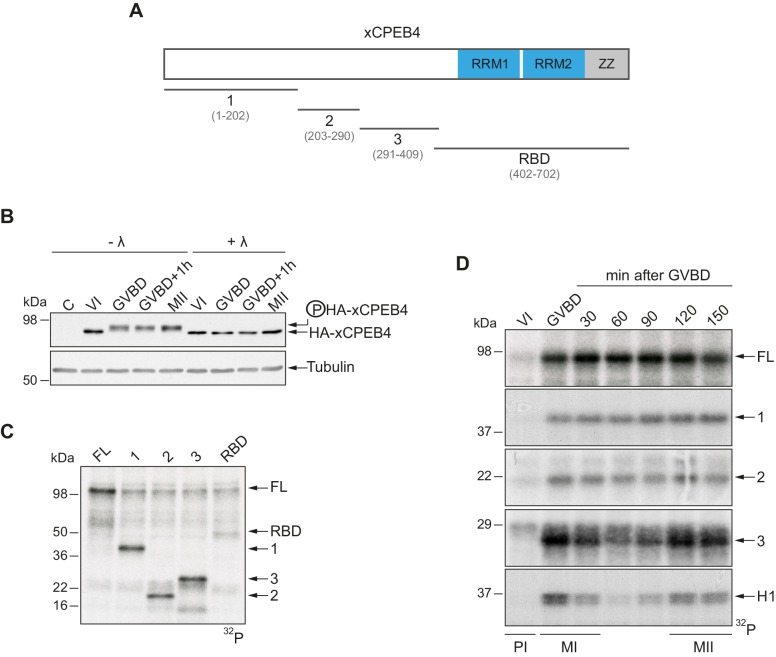
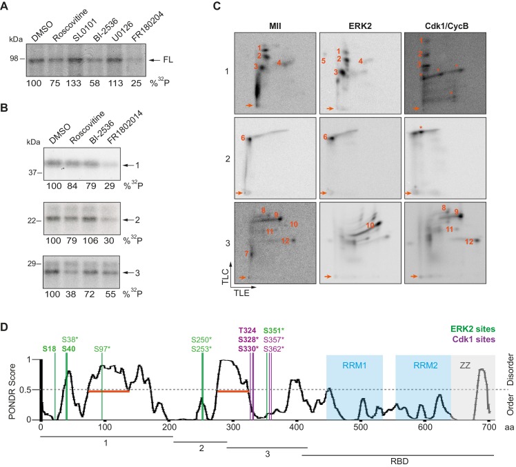
The four members of the vertebrate CPEB family of RNA-binding proteins share similar RNA-binding domains by which they regulate the translation of CPE-containing mRNAs, thereby controlling cell cycle and differentiation or synaptic plasticity. However, the N-terminal domains of CPEBs are distinct and contain specific regulatory post-translational modifications that presumably differentially integrate extracellular signals. Here we show that CPEB4 activity is regulated by ERK2- and Cdk1-mediated hyperphosphorylation. These phosphorylation events additively activate CPEB4 in M-phase by maintaining it in its monomeric state. In contrast, unphosphorylated CPEB4 phase separates into inactive, liquid-like droplets through its intrinsically disordered regions in the N-terminal domain. This dynamic and reversible regulation of CPEB4 is coordinated with that of CPEB1 through Cdk1, which inactivates CPEB1 while activating CPEB4, thereby integrating phase-specific signal transduction pathways to regulate cell cycle progression.

摘要

脊椎动物RNA结合蛋白CPEB家族的四个成员具有相似的RNA结合结构域,通过这些结构域它们调节含CPE的mRNA的翻译,从而控制细胞周期、分化或突触可塑性。然而,CPEB的N端结构域是不同的,并且包含特定的调节性翻译后修饰,这些修饰可能以不同方式整合细胞外信号。在此我们表明,CPEB4的活性受ERK2和Cdk1介导的过度磷酸化调节。这些磷酸化事件通过将CPEB4维持在单体状态,在M期累加激活CPEB4。相反,未磷酸化的CPEB4通过其N端结构域中固有无序区域相分离成无活性的液滴状。CPEB4这种动态且可逆的调节通过Cdk1与CPEB1的调节相协调,Cdk1使CPEB1失活同时激活CPEB4,从而整合阶段特异性信号转导途径以调节细胞周期进程。

https://cdn.ncbi.nlm.nih.gov/pmc/blobs/708b/5089860/344db7ed2a46/elife-19298-fig7.jpg
https://cdn.ncbi.nlm.nih.gov/pmc/blobs/708b/5089860/c308e0611ebc/elife-19298-fig1.jpg
https://cdn.ncbi.nlm.nih.gov/pmc/blobs/708b/5089860/ea0f6b6aa01c/elife-19298-fig1-figsupp1.jpg
https://cdn.ncbi.nlm.nih.gov/pmc/blobs/708b/5089860/dbc295a2777e/elife-19298-fig2.jpg
https://cdn.ncbi.nlm.nih.gov/pmc/blobs/708b/5089860/cf57651a3b5c/elife-19298-fig2-figsupp1.jpg
https://cdn.ncbi.nlm.nih.gov/pmc/blobs/708b/5089860/57490c7ce7b8/elife-19298-fig2-figsupp2.jpg
https://cdn.ncbi.nlm.nih.gov/pmc/blobs/708b/5089860/253ab00c812a/elife-19298-fig2-figsupp3.jpg
https://cdn.ncbi.nlm.nih.gov/pmc/blobs/708b/5089860/991c234e8cea/elife-19298-fig2-figsupp4.jpg
https://cdn.ncbi.nlm.nih.gov/pmc/blobs/708b/5089860/dea1c4cbba5b/elife-19298-fig3.jpg
https://cdn.ncbi.nlm.nih.gov/pmc/blobs/708b/5089860/4e64c847335e/elife-19298-fig3-figsupp1.jpg
https://cdn.ncbi.nlm.nih.gov/pmc/blobs/708b/5089860/0fe3d04dc490/elife-19298-fig3-figsupp2.jpg
https://cdn.ncbi.nlm.nih.gov/pmc/blobs/708b/5089860/681df3f3b8fa/elife-19298-fig3-figsupp3.jpg
https://cdn.ncbi.nlm.nih.gov/pmc/blobs/708b/5089860/7413434eb2ca/elife-19298-fig3-figsupp4.jpg
https://cdn.ncbi.nlm.nih.gov/pmc/blobs/708b/5089860/543d836d2836/elife-19298-fig4.jpg
https://cdn.ncbi.nlm.nih.gov/pmc/blobs/708b/5089860/21c3223bb255/elife-19298-fig4-figsupp1.jpg
https://cdn.ncbi.nlm.nih.gov/pmc/blobs/708b/5089860/dec7ce405842/elife-19298-fig4-figsupp2.jpg
https://cdn.ncbi.nlm.nih.gov/pmc/blobs/708b/5089860/1aa7a5808da8/elife-19298-fig5.jpg
https://cdn.ncbi.nlm.nih.gov/pmc/blobs/708b/5089860/10b4591c002c/elife-19298-fig5-figsupp1.jpg
https://cdn.ncbi.nlm.nih.gov/pmc/blobs/708b/5089860/7f3e47a96f65/elife-19298-fig6.jpg
https://cdn.ncbi.nlm.nih.gov/pmc/blobs/708b/5089860/22893d9f7eb1/elife-19298-fig6-figsupp1.jpg
https://cdn.ncbi.nlm.nih.gov/pmc/blobs/708b/5089860/d607d604b13b/elife-19298-fig6-figsupp2.jpg
https://cdn.ncbi.nlm.nih.gov/pmc/blobs/708b/5089860/344db7ed2a46/elife-19298-fig7.jpg

相似文献

1
CPEB4 is regulated during cell cycle by ERK2/Cdk1-mediated phosphorylation and its assembly into liquid-like droplets.CPEB4在细胞周期中受ERK2/Cdk1介导的磷酸化及其组装成液滴样结构的调控。
Elife. 2016 Nov 1;5:e19298. doi: 10.7554/eLife.19298.
2
XGef influences XRINGO/CDK1 signaling and CPEB activation during Xenopus oocyte maturation.XGef 影响 Xenopus oocyte 成熟过程中的 XRINGO/CDK1 信号和 CPEB 的激活。
Differentiation. 2011 Feb;81(2):133-40. doi: 10.1016/j.diff.2010.11.001. Epub 2010 Dec 8.
3
Meiosis requires a translational positive loop where CPEB1 ensues its replacement by CPEB4.减数分裂需要一个翻译正反馈环,在此环中 CPEB1 被 CPEB4 取代。
EMBO J. 2010 Jul 7;29(13):2182-93. doi: 10.1038/emboj.2010.111. Epub 2010 Jun 8.
4
Mitotic cell-cycle progression is regulated by CPEB1 and CPEB4-dependent translational control.有丝分裂细胞周期进程受 CPEB1 和 CPEB4 依赖性翻译控制的调节。
Nat Cell Biol. 2010 May;12(5):447-56. doi: 10.1038/ncb2046. Epub 2010 Apr 4.
5
Differential mRNA translation and meiotic progression require Cdc2-mediated CPEB destruction.差异性mRNA翻译和减数分裂进程需要Cdc2介导的CPEB破坏。
EMBO J. 2002 Apr 2;21(7):1833-44. doi: 10.1093/emboj/21.7.1833.
6
RINGO/cdk1 and CPEB mediate poly(A) tail stabilization and translational regulation by ePAB.RINGO/cdk1和CPEB通过ePAB介导多聚腺苷酸(poly(A))尾的稳定和翻译调控。
Genes Dev. 2007 Oct 15;21(20):2571-9. doi: 10.1101/gad.1593007.
7
Sequential Functions of CPEB1 and CPEB4 Regulate Pathologic Expression of Vascular Endothelial Growth Factor and Angiogenesis in Chronic Liver Disease.CPEB1 和 CPEB4 的级联功能调节慢性肝病中血管内皮生长因子的病理性表达和血管生成。
Gastroenterology. 2016 Apr;150(4):982-97.e30. doi: 10.1053/j.gastro.2015.11.038. Epub 2015 Nov 26.
8
Vg1RBP phosphorylation by Erk2 MAP kinase correlates with the cortical release of Vg1 mRNA during meiotic maturation of Xenopus oocytes.在非洲爪蟾卵母细胞减数分裂成熟过程中,Erk2丝裂原活化蛋白激酶对Vg1RBP的磷酸化作用与Vg1 mRNA的皮质释放相关。
RNA. 2009 Jun;15(6):1121-33. doi: 10.1261/rna.1195709. Epub 2009 Apr 17.
9
Possible involvement of Nemo-like kinase 1 in Xenopus oocyte maturation as a kinase responsible for Pumilio1, Pumilio2, and CPEB phosphorylation.可能涉及 Nemo 样激酶 1 参与非洲爪蟾卵母细胞成熟,作为负责 Pumilio1、Pumilio2 和 CPEB 磷酸化的激酶。
Biochemistry. 2011 Jun 28;50(25):5648-59. doi: 10.1021/bi2002696. Epub 2011 Jun 4.
10
CDK1 and calcineurin regulate Maskin association with eIF4E and translational control of cell cycle progression.细胞周期蛋白依赖性激酶1(CDK1)和钙调神经磷酸酶调节Maskin与真核生物翻译起始因子4E(eIF4E)的结合以及细胞周期进程的翻译控制。
Nat Struct Mol Biol. 2006 Dec;13(12):1128-34. doi: 10.1038/nsmb1169. Epub 2006 Nov 5.

引用本文的文献

1
Role of CPEBs in Learning and Memory.CPEB 在学习与记忆中的作用。
J Neurochem. 2025 Sep;169(9):e70226. doi: 10.1111/jnc.70226.
2
Epigenetic Mechanisms Shaping Spine Regulation: Unveiling the Role of Cytoskeletal Dynamics and Localized Protein Synthesis.塑造脊柱调节的表观遗传机制:揭示细胞骨架动力学和局部蛋白质合成的作用
Mol Neurobiol. 2025 Jun 3. doi: 10.1007/s12035-025-05045-7.
3
CPEB4 modulates liver cancer progression by translationally regulating hepcidin expression and sensitivity to ferroptosis.CPEB4通过翻译调控铁调素表达和对铁死亡的敏感性来调节肝癌进展。

本文引用的文献

1
Amyloidogenic Oligomerization Transforms Drosophila Orb2 from a Translation Repressor to an Activator.淀粉样寡聚化将果蝇Orb2从翻译抑制因子转变为激活因子。
Cell. 2015 Dec 3;163(6):1468-83. doi: 10.1016/j.cell.2015.11.020.
2
Formation and Maturation of Phase-Separated Liquid Droplets by RNA-Binding Proteins.RNA结合蛋白介导的相分离液滴的形成与成熟
Mol Cell. 2015 Oct 15;60(2):208-19. doi: 10.1016/j.molcel.2015.08.018. Epub 2015 Sep 24.
3
Phase separation by low complexity domains promotes stress granule assembly and drives pathological fibrillization.
JHEP Rep. 2024 Dec 12;7(3):101296. doi: 10.1016/j.jhepr.2024.101296. eCollection 2025 Mar.
4
Cytoplasmic regulation of the poly(A) tail length as a potential therapeutic target.聚腺苷酸尾长度的细胞质调控作为一种潜在的治疗靶点。
RNA. 2025 Feb 19;31(3):402-415. doi: 10.1261/rna.080333.124.
5
Mis-splicing of a neuronal microexon promotes CPEB4 aggregation in ASD.神经元微小外显子的错误剪接促进了自闭症谱系障碍中CPEB4的聚集。
Nature. 2025 Jan;637(8045):496-503. doi: 10.1038/s41586-024-08289-w. Epub 2024 Dec 4.
6
NPPC and AREG supplementation in IVM systems alter mRNA translation and decay programs-related gene expression in bovine COC.在体外成熟(IVM)系统中补充尿钠肽前体C(NPPC)和双调蛋白(AREG)会改变牛卵丘卵母细胞复合体(COC)中与mRNA翻译和衰变程序相关的基因表达。
Anim Reprod. 2024 Jul 8;21(2):e20230101. doi: 10.1590/1984-3143-AR2023-0101. eCollection 2024.
7
Core-shell model of the clusters of CPEB4 isoforms preceding liquid-liquid phase separation.CPEB4 异构体簇液-液相分离前的核壳模型。
Biophys J. 2024 Aug 20;123(16):2604-2622. doi: 10.1016/j.bpj.2024.06.027. Epub 2024 Jun 28.
8
Physiology and pharmacological targeting of phase separation.相分离的生理学和药理学靶向。
J Biomed Sci. 2024 Jan 20;31(1):11. doi: 10.1186/s12929-024-00993-z.
9
RNA-Binding Proteins as Critical Post-Transcriptional Regulators of Cardiac Regeneration.RNA结合蛋白作为心脏再生的关键转录后调节因子
Int J Mol Sci. 2023 Jul 26;24(15):12004. doi: 10.3390/ijms241512004.
10
CPEB and translational control by cytoplasmic polyadenylation: impact on synaptic plasticity, learning, and memory.CPEB 和细胞质多聚腺苷酸化的翻译控制:对突触可塑性、学习和记忆的影响。
Mol Psychiatry. 2023 Jul;28(7):2728-2736. doi: 10.1038/s41380-023-02088-x. Epub 2023 May 2.
低复杂性结构域介导的相分离促进应激颗粒组装并驱动病理性纤维化。
Cell. 2015 Sep 24;163(1):123-33. doi: 10.1016/j.cell.2015.09.015.
4
Global Analysis of CPEBs Reveals Sequential and Non-Redundant Functions in Mitotic Cell Cycle.CPEB的全局分析揭示了有丝分裂细胞周期中的顺序性和非冗余功能。
PLoS One. 2015 Sep 23;10(9):e0138794. doi: 10.1371/journal.pone.0138794. eCollection 2015.
5
A Liquid-to-Solid Phase Transition of the ALS Protein FUS Accelerated by Disease Mutation.疾病突变加速 ALS 蛋白 FUS 的液-固相变。
Cell. 2015 Aug 27;162(5):1066-77. doi: 10.1016/j.cell.2015.07.047.
6
SUMOylation Is an Inhibitory Constraint that Regulates the Prion-like Aggregation and Activity of CPEB3.小泛素样修饰是一种调节CPEB3的朊病毒样聚集和活性的抑制性限制因素。
Cell Rep. 2015 Jun 23;11(11):1694-702. doi: 10.1016/j.celrep.2015.04.061. Epub 2015 Jun 11.
7
Phase transition of a disordered nuage protein generates environmentally responsive membraneless organelles.无序云状蛋白的相变产生环境响应性无膜细胞器。
Mol Cell. 2015 Mar 5;57(5):936-947. doi: 10.1016/j.molcel.2015.01.013.
8
Folding of an intrinsically disordered protein by phosphorylation as a regulatory switch.磷酸化诱导无规卷曲蛋白折叠作为一种调控开关。
Nature. 2015 Mar 5;519(7541):106-9. doi: 10.1038/nature13999. Epub 2014 Dec 22.
9
Ultrasensitivity part II: multisite phosphorylation, stoichiometric inhibitors, and positive feedback.超敏感性第二部分:多位点磷酸化、化学计量抑制剂与正反馈
Trends Biochem Sci. 2014 Nov;39(11):556-69. doi: 10.1016/j.tibs.2014.09.003. Epub 2014 Oct 23.
10
Arsenite-activated JNK signaling enhances CPEB4-Vinexin interaction to facilitate stress granule assembly and cell survival.亚砷酸盐激活的JNK信号增强CPEB4与Vinexin的相互作用,以促进应激颗粒组装和细胞存活。
PLoS One. 2014 Sep 19;9(9):e107961. doi: 10.1371/journal.pone.0107961. eCollection 2014.