Fluidigm Canada Inc., 1380 Rodick Road, Markham, Ontario L3R 4G5, Canada.
Department of Pathology, Hospital for Sick Children, 555 University Avenue, Ontario M5G 1X8, Canada.
Sci Rep. 2016 Nov 4;6:36641. doi: 10.1038/srep36641.
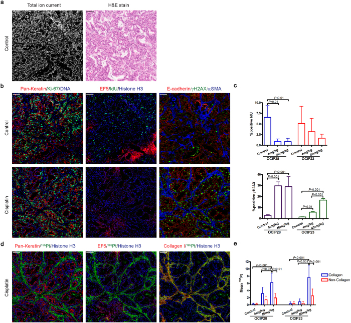
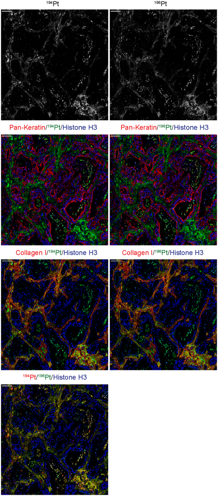
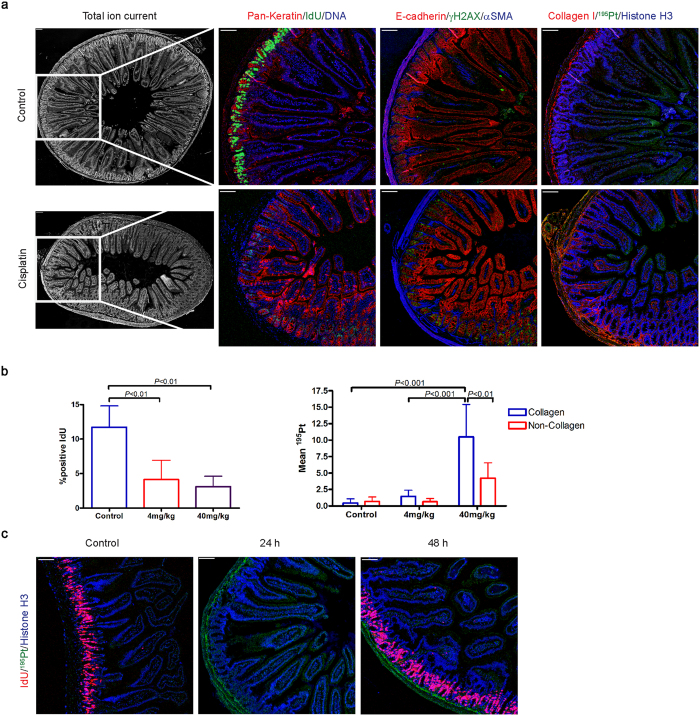
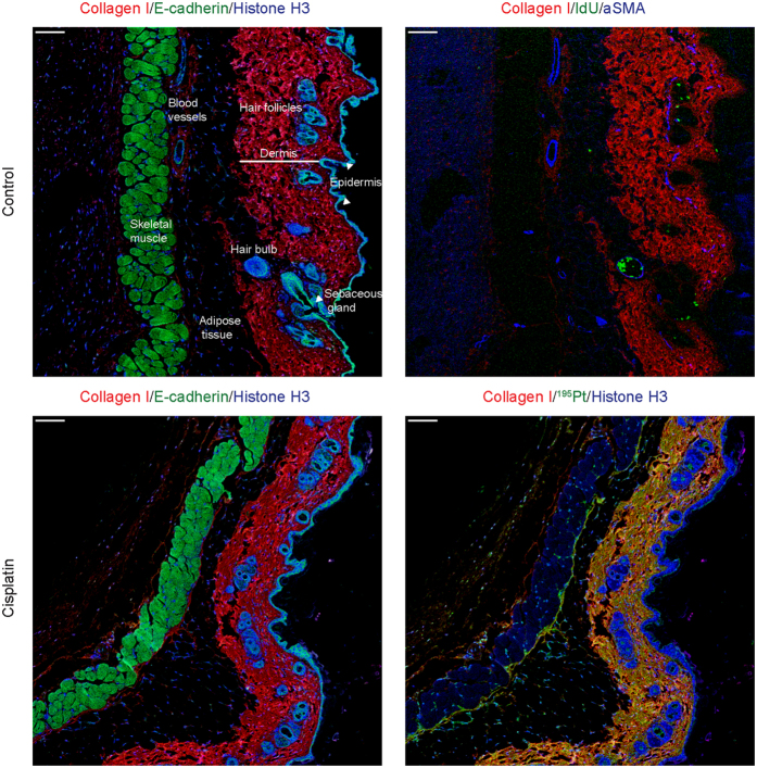
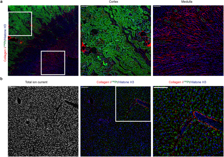
Imaging mass cytometry was used for direct visualization of platinum localization in tissue sections from tumor and normal tissues of cisplatin-treated mice bearing pancreas cancer patient-derived xenografts. This recently-developed technology enabled simultaneous detection of multiple markers to define cell lineage, DNA damage response, cell proliferation and functional state, providing a highly detailed view of drug incorporation in tumor and normal tissues at the cellular level. A striking and unanticipated finding was the extensive binding of platinum to collagen fibers in both tumor and normal mouse tissues. Time course experiments indicated the slow release of stroma-bound platinum, although it is currently unclear if released platinum retains biological activity. Imaging mass cytometry offers a unique window into the in vivo effects of platinum compounds, and it is likely that this can be extended into the clinic in order to optimize the use of this important class of agent.
成像质谱细胞术被用于直接观察顺铂处理的胰腺癌患者来源异种移植小鼠的肿瘤和正常组织切片中的铂定位。这项最近开发的技术能够同时检测多个标志物来定义细胞谱系、DNA 损伤反应、细胞增殖和功能状态,从而在细胞水平上提供对肿瘤和正常组织中药物掺入的高度详细视图。一个惊人且出乎意料的发现是铂在肿瘤和正常小鼠组织中的胶原纤维上的广泛结合。时程实验表明,基质结合的铂释放缓慢,尽管目前尚不清楚释放的铂是否保留生物活性。成像质谱细胞术为研究铂化合物的体内作用提供了一个独特的窗口,并且很可能将其扩展到临床,以优化这类重要药物的使用。