• 文献检索
  • 文档翻译
  • 深度研究
  • 学术资讯
  • Suppr Zotero 插件Zotero 插件
  • 邀请有礼
  • 套餐&价格
  • 历史记录
应用&插件
Suppr Zotero 插件Zotero 插件浏览器插件Mac 客户端Windows 客户端微信小程序
定价
高级版会员购买积分包购买API积分包
服务
文献检索文档翻译深度研究API 文档MCP 服务
关于我们
关于 Suppr公司介绍联系我们用户协议隐私条款
关注我们

Suppr 超能文献

核心技术专利:CN118964589B侵权必究
粤ICP备2023148730 号-1Suppr @ 2026

文献检索

告别复杂PubMed语法,用中文像聊天一样搜索,搜遍4000万医学文献。AI智能推荐,让科研检索更轻松。

立即免费搜索

文件翻译

保留排版,准确专业,支持PDF/Word/PPT等文件格式,支持 12+语言互译。

免费翻译文档

深度研究

AI帮你快速写综述,25分钟生成高质量综述,智能提取关键信息,辅助科研写作。

立即免费体验

奥沙利铂诱导化疗引起的局灶性窦性阻塞综合征:一例报告

Focal Sinusoidal Obstruction Syndrome Caused by Oxaliplatin-Induced Chemotherapy: A Case Report.

作者信息

Kawai Takaharu, Yamazaki Shintaro, Iwama Atsuko, Higaki Tokio, Sugitani Masahiko, Takayama Tadatoshi

机构信息

Department of Digestive Surgery and Pathology, School of Medicine, Nihon University, Tokyo, Japan.

出版信息

Hepat Mon. 2016 Aug 21;16(9):e37572. doi: 10.5812/hepatmon.37572. eCollection 2016 Sep.

DOI:10.5812/hepatmon.37572
PMID:27822263
原文链接:https://pmc.ncbi.nlm.nih.gov/articles/PMC5091096/
Abstract

INTRODUCTION

Sinusoidal obstruction syndrome (SOS) is a severe adverse event of long-term chemotherapy in patients with colorectal cancer. It usually develops as liver congestion due to diffuse microscopic obstruction in liver parenchyma. In contrast, it sometimes appears as a liver mass occurring with local parenchymal hemorrhaging, and is often misdiagnosed as liver metastasis.

CASE PRESENTATION

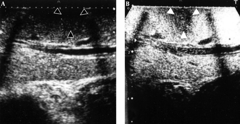
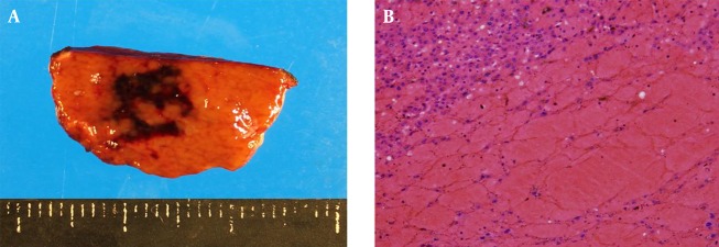
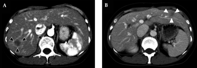
A 40-year-old woman with rectal cancer underwent high anterior resection and partial liver resection of segment 7 due to synchronous liver metastasis. She received oxaliplatin-based chemotherapy (mFOLFOX6) as adjuvant chemotherapy for 6 months. A 13-mm irregular low-echoic mass was detected by CT in segment 3 of the liver 12 months after the operation. The mass was again resected as a liver metastasis because it had increased in size. The pathological diagnosis was focal SOS, which showed sinusoidal dilation and congestion by hepatocyte trabeculae in the liver parenchyma.

CONCLUSIONS

Atypical irregular tumors should be considered as SOS when the patient has received oxaliplatin-based chemotherapy. A qualitative imaging modality diagnosis, such as with diffusion-weighted MRI, is superior to a morphological diagnosis in focal SOS. This imaging modality can prevent unnecessary operations.

摘要

引言

窦性阻塞综合征(SOS)是结直肠癌患者长期化疗的严重不良事件。它通常是由于肝实质内弥漫性微小阻塞导致肝脏充血而发展形成。相比之下,它有时表现为伴有局部实质出血的肝脏肿块,且常被误诊为肝转移。

病例报告

一名40岁的直肠癌女性患者因同时性肝转移接受了高位前切除术和肝7段部分切除术。她接受了以奥沙利铂为基础的化疗(mFOLFOX6)作为辅助化疗,为期6个月。术后12个月,CT在肝脏3段检测到一个13毫米不规则低回声肿块。由于肿块增大,再次将其作为肝转移瘤切除。病理诊断为局灶性SOS,显示肝实质内肝细胞小梁间的窦性扩张和充血。

结论

当患者接受以奥沙利铂为基础的化疗时,非典型不规则肿瘤应考虑为SOS。定性成像方式诊断,如扩散加权磁共振成像,在局灶性SOS中优于形态学诊断。这种成像方式可以避免不必要的手术。

https://cdn.ncbi.nlm.nih.gov/pmc/blobs/7a4e/5091096/e0c03196f028/hepatmon-16-09-37572-g004.jpg
https://cdn.ncbi.nlm.nih.gov/pmc/blobs/7a4e/5091096/600e44beded0/hepatmon-16-09-37572-g001.jpg
https://cdn.ncbi.nlm.nih.gov/pmc/blobs/7a4e/5091096/1640e5726279/hepatmon-16-09-37572-g002.jpg
https://cdn.ncbi.nlm.nih.gov/pmc/blobs/7a4e/5091096/fdb132e04d1e/hepatmon-16-09-37572-g003.jpg
https://cdn.ncbi.nlm.nih.gov/pmc/blobs/7a4e/5091096/e0c03196f028/hepatmon-16-09-37572-g004.jpg

相似文献

1
Focal Sinusoidal Obstruction Syndrome Caused by Oxaliplatin-Induced Chemotherapy: A Case Report.奥沙利铂诱导化疗引起的局灶性窦性阻塞综合征:一例报告
Hepat Mon. 2016 Aug 21;16(9):e37572. doi: 10.5812/hepatmon.37572. eCollection 2016 Sep.
2
Oxaliplatin-induced sinusoidal obstruction syndrome mimicking metastatic colon cancer in the liver.奥沙利铂诱导的肝窦阻塞综合征酷似肝脏转移性结肠癌。
Oncol Lett. 2016 Apr;11(4):2861-2864. doi: 10.3892/ol.2016.4286. Epub 2016 Feb 29.
3
Oxaliplatin-related sinusoidal obstruction syndrome mimicking metastatic liver tumors.奥沙利铂相关的窦状隙阻塞综合征酷似转移性肝肿瘤。
Hepatol Res. 2013 Jun;43(6):685-9. doi: 10.1111/j.1872-034X.2012.01114.x.
4
Oxaliplatin-Induced Sinusoidal Obstruction Syndrome Masquerading as Colorectal Liver Metastasis: A Case Report.奥沙利铂诱导的肝窦阻塞综合征伪装为结直肠癌肝转移:一例报告。
J Gastrointest Cancer. 2023 Jun;54(2):682-686. doi: 10.1007/s12029-022-00835-x. Epub 2022 Jun 6.
5
Autotaxin is a novel surrogate marker for oxaliplatin-related sinusoidal obstruction syndrome in patients with colorectal liver metastasis.自分泌运动因子是结直肠癌肝转移患者中奥沙利铂相关肝窦阻塞综合征的一种新型替代标志物。
Hepatol Res. 2023 Aug;53(8):761-770. doi: 10.1111/hepr.13901. Epub 2023 Apr 2.
6
Chemotherapy-induced Sinusoidal Injury (CSI) score: a novel histologic assessment of chemotherapy-related hepatic sinusoidal injury in patients with colorectal liver metastasis.化疗诱导的窦性损伤(CSI)评分:一种对结直肠癌肝转移患者化疗相关肝窦性损伤的新型组织学评估。
BMC Cancer. 2017 Jan 7;17(1):35. doi: 10.1186/s12885-016-2998-2.
7
Oxaliplatin toxicity presenting as a liver nodule - case report.以肝结节表现的奥沙利铂毒性——病例报告
BMC Cancer. 2015 Apr 10;15:247. doi: 10.1186/s12885-015-1247-4.
8
Oxaliplatin-induced hepatic sinusoidal obstruction syndrome in a patient with gastric cancer: A case report.奥沙利铂诱发胃癌患者肝窦阻塞综合征:一例报告
Mol Clin Oncol. 2018 Mar;8(3):453-456. doi: 10.3892/mco.2017.1540. Epub 2017 Dec 18.
9
Correlation between Gd-EOB-DTPA-enhanced MR imaging findings and OATP1B3 expression in chemotherapy-associated sinusoidal obstruction syndrome.钆塞酸二钠增强磁共振成像表现与化疗相关肝窦阻塞综合征中有机阴离子转运多肽1B3表达的相关性
Abdom Imaging. 2015 Oct;40(8):3099-103. doi: 10.1007/s00261-015-0503-z.
10
Diagnostic performance of contrast-enhanced CT-scan in sinusoidal obstruction syndrome induced by chemotherapy of colorectal liver metastases: Radio-pathological correlation.对比增强 CT 扫描对结直肠肝转移化疗后引起的肝窦阻塞综合征的诊断性能:放射病理学相关性。
Eur J Radiol. 2017 Sep;94:180-190. doi: 10.1016/j.ejrad.2017.06.025. Epub 2017 Jul 3.

引用本文的文献

1
Oxaliplatin-Induced Sinusoidal Obstruction Syndrome Masquerading as Colorectal Liver Metastasis: A Case Report.奥沙利铂诱导的肝窦阻塞综合征伪装为结直肠癌肝转移:一例报告。
J Gastrointest Cancer. 2023 Jun;54(2):682-686. doi: 10.1007/s12029-022-00835-x. Epub 2022 Jun 6.
2
FDG-PET/CT imaging findings of hepatic tumors and tumor-like lesions based on molecular background.基于分子背景的肝脏肿瘤和肿瘤样病变的 FDG-PET/CT 影像学表现。
Jpn J Radiol. 2020 Aug;38(8):697-718. doi: 10.1007/s11604-020-00961-1. Epub 2020 Apr 3.
3
Noninvasive imaging diagnosis of sinusoidal obstruction syndrome: a pictorial review.

本文引用的文献

1
Oxaliplatin toxicity presenting as a liver nodule - case report.以肝结节表现的奥沙利铂毒性——病例报告
BMC Cancer. 2015 Apr 10;15:247. doi: 10.1186/s12885-015-1247-4.
2
Oxaliplatin-induced liver injury mimicking metastatic tumor on images: a case report.奥沙利铂诱导的肝损伤模拟影像学转移瘤:病例报告。
Jpn J Clin Oncol. 2013 Oct;43(10):1034-8. doi: 10.1093/jjco/hyt113. Epub 2013 Aug 19.
3
Oxaliplatin-related sinusoidal obstruction syndrome mimicking metastatic liver tumors.奥沙利铂相关的窦状隙阻塞综合征酷似转移性肝肿瘤。
窦性阻塞综合征的无创成像诊断:图文综述
Insights Imaging. 2019 Nov 20;10(1):110. doi: 10.1186/s13244-019-0791-x.
4
Sinusoidal obstruction syndrome: A systematic review of etiologies, clinical symptoms, and magnetic resonance imaging features.窦性阻塞综合征:病因、临床症状及磁共振成像特征的系统评价
World J Clin Cases. 2019 Sep 26;7(18):2746-2759. doi: 10.12998/wjcc.v7.i18.2746.
5
Histopathological and biochemical assessment of liver damage in albino Wistar rats treated with cytotoxic platinum compounds in combination with 5-fluorouracil.对白化Wistar大鼠使用细胞毒性铂化合物与5-氟尿嘧啶联合治疗后肝脏损伤的组织病理学和生化评估。
Arch Med Sci. 2019 Jul;15(4):1092-1103. doi: 10.5114/aoms.2019.86064. Epub 2019 Jun 20.
6
Diagnostic value/performance of radiological liver imaging during chemoterapy for gastrointestinal malignancy: a critical review.胃肠道恶性肿瘤化疗期间肝脏影像学检查的诊断价值/效能:一项批判性综述
Acta Biomed. 2019 Apr 24;90(5-S):51-61. doi: 10.23750/abm.v90i5-S.8346.
Hepatol Res. 2013 Jun;43(6):685-9. doi: 10.1111/j.1872-034X.2012.01114.x.
4
A nomogram predicting disease-free survival in patients with colorectal liver metastases treated with hepatic resection: multicenter data collection as a Project Study for Hepatic Surgery of the Japanese Society of Hepato-Biliary-Pancreatic Surgery.预测接受肝切除术治疗结直肠肝转移患者无病生存的列线图:日本肝胆胰外科学会肝脏外科研讨会项目的多中心数据收集。
J Hepatobiliary Pancreat Sci. 2012 Jan;19(1):72-84. doi: 10.1007/s00534-011-0460-z.
5
Value of PET/CT in the management of liver metastases, part 1.PET/CT 在肝转移瘤管理中的价值,第 1 部分。
AJR Am J Roentgenol. 2011 Aug;197(2):W256-9. doi: 10.2214/AJR.10.6331.
6
Predicting high grade lesions of sinusoidal obstruction syndrome related to oxaliplatin-based chemotherapy for colorectal liver metastases: correlation with post-hepatectomy outcome.预测结直肠肝转移瘤患者接受奥沙利铂为基础的化疗后发生肝窦阻塞综合征高分级病变:与肝切除术后结局的相关性。
Ann Surg. 2010 Mar;251(3):454-60. doi: 10.1097/SLA.0b013e3181c79403.
7
Perioperative chemotherapy with FOLFOX4 and surgery versus surgery alone for resectable liver metastases from colorectal cancer (EORTC Intergroup trial 40983): a randomised controlled trial.FOLFOX4围手术期化疗联合手术与单纯手术治疗可切除的结直肠癌肝转移(欧洲癌症研究与治疗组织(EORTC)国际协作组试验40983):一项随机对照试验
Lancet. 2008 Mar 22;371(9617):1007-16. doi: 10.1016/S0140-6736(08)60455-9.
8
Focal liver masses: characterization with diffusion-weighted echo-planar MR imaging.肝脏局灶性肿块:磁共振扩散加权回波平面成像特征分析
Radiology. 1997 Sep;204(3):739-44. doi: 10.1148/radiology.204.3.9280252.
9
The natural history of primary and secondary malignant tumors of the liver. I. The prognosis for patients with hepatic metastases from colonic and rectal carcinoma by laparotomy.肝脏原发性和继发性恶性肿瘤的自然病史。一、经剖腹手术治疗的结肠和直肠癌肝转移患者的预后。
Cancer. 1969 Jan;23(1):198-202. doi: 10.1002/1097-0142(196901)23:1<198::aid-cncr2820230126>3.0.co;2-j.
10
Liver metastases found by follow-up of patients operated on for colorectal cancer.
Cancer. 1976 Mar;37(3):1454-7. doi: 10.1002/1097-0142(197603)37:3<1454::aid-cncr2820370329>3.0.co;2-d.