Metlapally Ravikanth, Park Han Na, Chakraborty Ranjay, Wang Kevin K, Tan Christopher C, Light Jacob G, Pardue Machelle T, Wildsoet Christine F
School of Optometry, University of California at Berkeley, Berkeley, California, United States.
Department of Ophthalmology at Emory University, Atlanta, Georgia, United States.
Invest Ophthalmol Vis Sci. 2016 Nov 1;57(14):6089-6097. doi: 10.1167/iovs.16-19563.
MicroRNA (miRNAs) have been previously implicated in scleral remodeling in normal eye growth. They have the potential to be therapeutic targets for prevention/retardation of exaggerated eye growth in myopia by modulating scleral matrix remodeling. To explore this potential, genome-wide miRNA and messenger RNA (mRNA) scleral profiles in myopic and control eyes from mice were studied.
C57BL/6J mice (n = 7; P28) reared under a 12L:12D cycle were form-deprived (FD) unilaterally for 2 weeks. Refractive error and axial length changes were measured using photorefraction and 1310-nm spectral-domain optical coherence tomography, respectively. Scleral RNA samples from FD and fellow control eyes were processed for microarray assay. Statistical analyses were performed using National Institute of Aging array analysis tool; group comparisons were made using ANOVA, and gene ontologies were identified using software available on the Web. Findings were confirmed using quantitative PCR in a separate group of mice (n = 7).
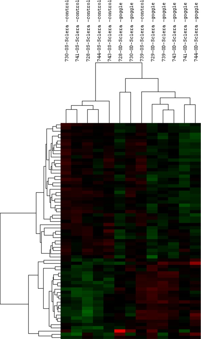
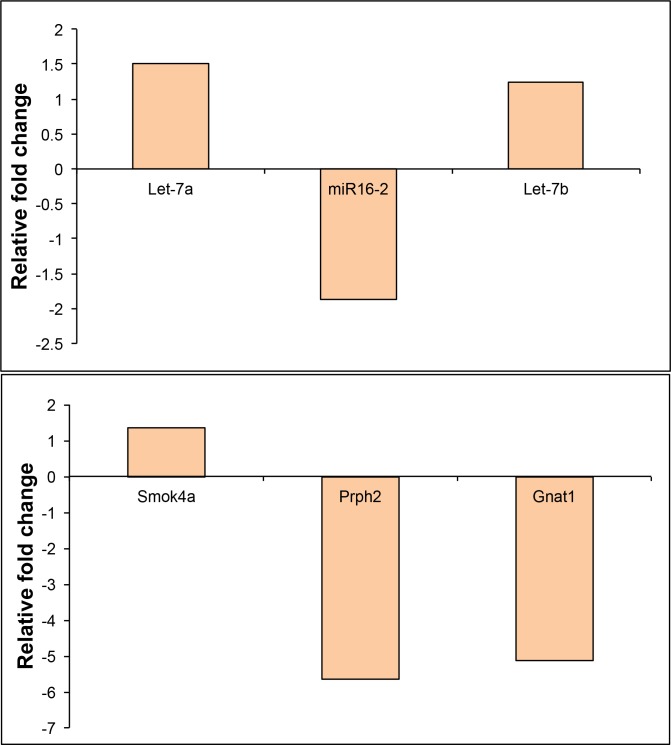
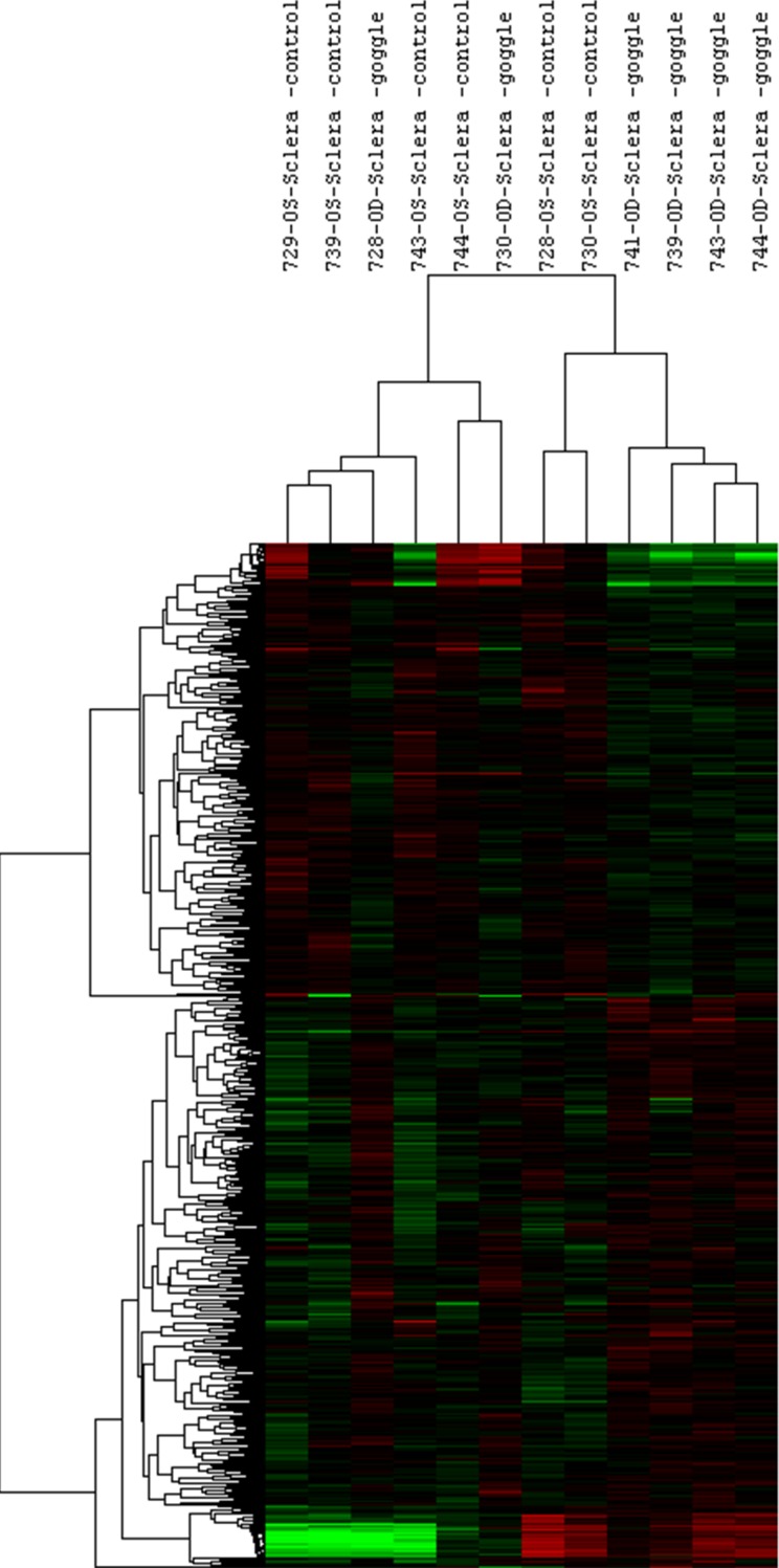
Form-deprived eyes showed myopic shifts in refractive error (-2.02 ± 0.47 D; P < 0.01). Comparison of the scleral RNA profiles of test eyes with those of control eyes revealed 54 differentially expressed miRNAs and 261 mRNAs fold-change >1.25 (maximum fold change = 1.63 and 2.7 for miRNAs and mRNAs, respectively) (P < 0.05; minimum, P = 0.0001). Significant ontologies showing gene over-representation (P < 0.05) included intermediate filament organization, scaffold protein binding, detection of stimuli, calcium ion, G protein, and phototransduction. Significant differential expression of Let-7a and miR-16-2, and Smok4a, Prph2, and Gnat1 were confirmed.
Scleral mi- and mRNAs showed differential expression linked to myopia, supporting the involvement of miRNAs in eye growth regulation. The observed general trend of relatively small fold-changes suggests a tightly controlled, regulatory mechanism for scleral gene expression.
微小RNA(miRNA)此前已被证明参与正常眼球生长过程中的巩膜重塑。它们有可能通过调节巩膜基质重塑成为预防/延缓近视中眼球过度生长的治疗靶点。为了探索这种潜力,我们研究了小鼠近视和对照眼中全基因组的miRNA和信使核糖核酸(mRNA)巩膜谱。
将饲养在12小时光照:12小时黑暗周期下的C57BL/6J小鼠(n = 7;出生后28天)单眼形觉剥夺(FD)2周。分别使用 photorefraction和1310纳米光谱域光学相干断层扫描测量屈光不正和眼轴长度变化。对FD眼和对侧对照眼的巩膜RNA样本进行微阵列分析。使用美国国立衰老研究所阵列分析工具进行统计分析;采用方差分析进行组间比较,并使用网络上可用的软件识别基因本体。在另一组小鼠(n = 7)中使用定量PCR对结果进行验证。
形觉剥夺眼的屈光不正出现近视性偏移(-2.02 ± 0.47 D;P < 0.01)。将试验眼与对照眼的巩膜RNA谱进行比较,发现54种差异表达的miRNA和261种mRNA的变化倍数>1.25(miRNA和mRNA的最大变化倍数分别为1.63和2.7)(P < 0.05;最小值,P = 0.0001)。显示基因过度表达(P < 0.05)的显著本体包括中间丝组织、支架蛋白结合、刺激检测、钙离子、G蛋白和光转导。Let-7a和miR-16-2以及Smok4a、Prph2和Gnat1的显著差异表达得到了验证。
巩膜miRNA和mRNA显示出与近视相关的差异表达,支持miRNA参与眼生长调节。观察到的相对较小变化倍数的总体趋势表明巩膜基因表达存在严格控制的调节机制。