• 文献检索
  • 文档翻译
  • 深度研究
  • 学术资讯
  • Suppr Zotero 插件Zotero 插件
  • 邀请有礼
  • 套餐&价格
  • 历史记录
应用&插件
Suppr Zotero 插件Zotero 插件浏览器插件Mac 客户端Windows 客户端微信小程序
定价
高级版会员购买积分包购买API积分包
服务
文献检索文档翻译深度研究API 文档MCP 服务
关于我们
关于 Suppr公司介绍联系我们用户协议隐私条款
关注我们

Suppr 超能文献

核心技术专利:CN118964589B侵权必究
粤ICP备2023148730 号-1Suppr @ 2026

文献检索

告别复杂PubMed语法,用中文像聊天一样搜索,搜遍4000万医学文献。AI智能推荐,让科研检索更轻松。

立即免费搜索

文件翻译

保留排版,准确专业,支持PDF/Word/PPT等文件格式,支持 12+语言互译。

免费翻译文档

深度研究

AI帮你快速写综述,25分钟生成高质量综述,智能提取关键信息,辅助科研写作。

立即免费体验

与心肌细胞电耦合的肌成纤维细胞改变传导:来自详细组织结构模型的细胞水平见解

Myofibroblasts Electrotonically Coupled to Cardiomyocytes Alter Conduction: Insights at the Cellular Level from a Detailed Tissue Structure Model.

作者信息

Jousset Florian, Maguy Ange, Rohr Stephan, Kucera Jan P

机构信息

Department of Physiology, University of Bern Bern, Switzerland.

出版信息

Front Physiol. 2016 Oct 27;7:496. doi: 10.3389/fphys.2016.00496. eCollection 2016.

DOI:10.3389/fphys.2016.00496
PMID:27833567
原文链接:https://pmc.ncbi.nlm.nih.gov/articles/PMC5081362/
Abstract

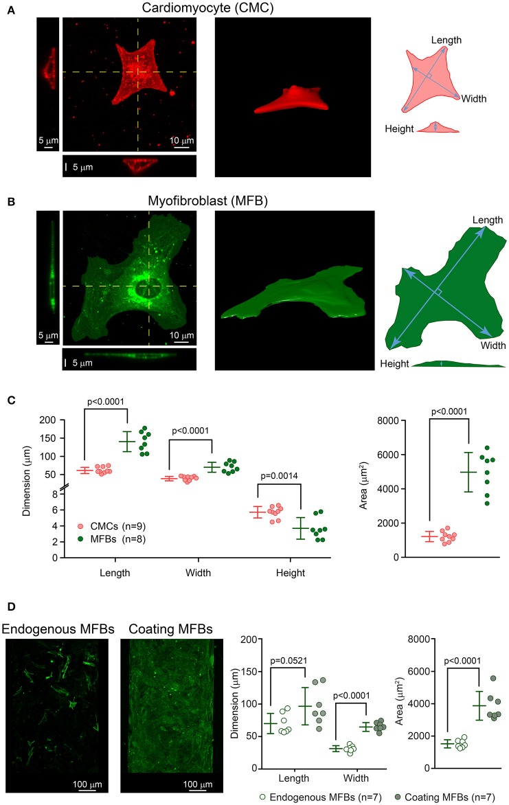
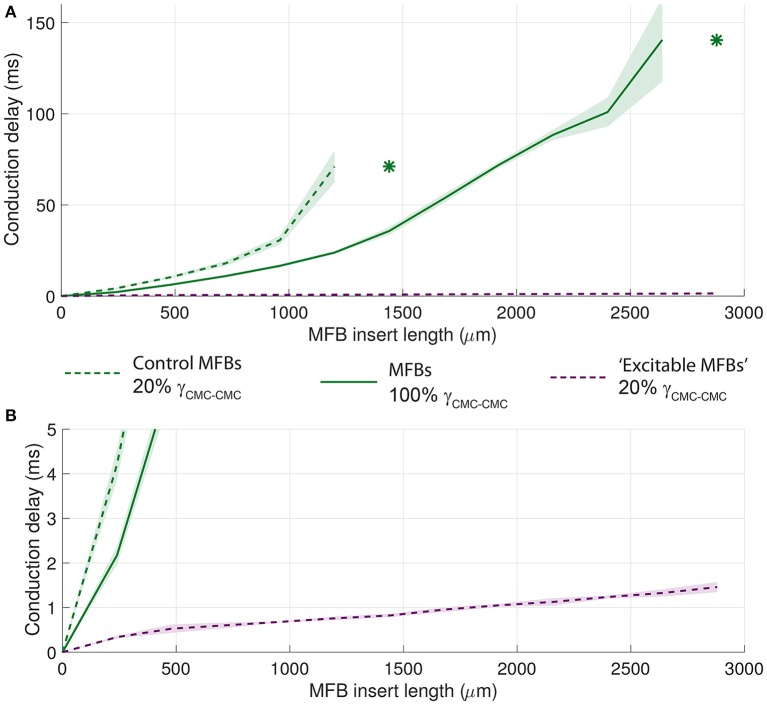
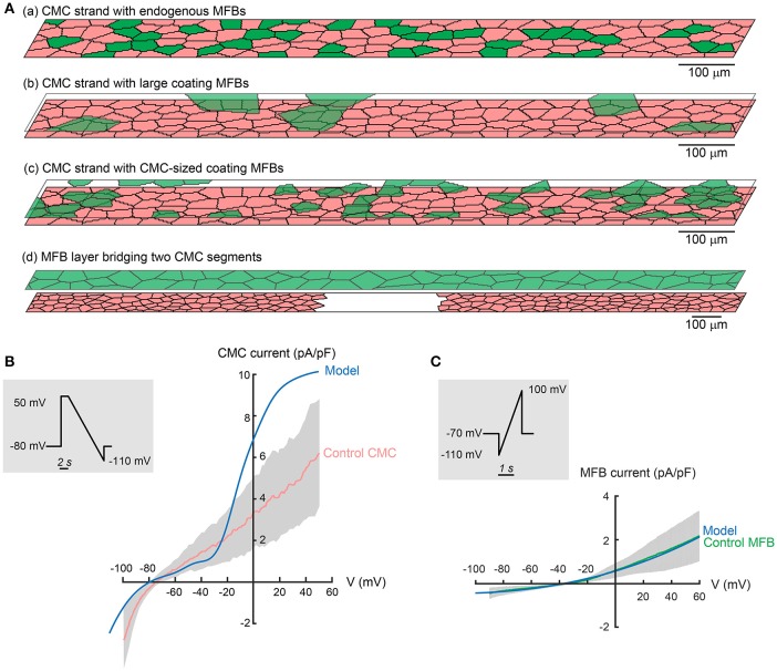
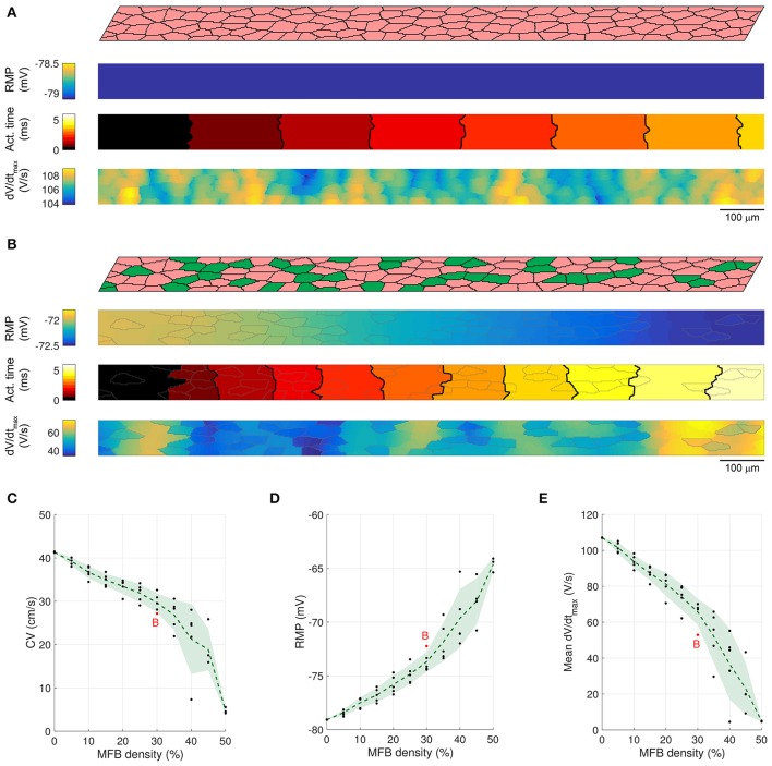
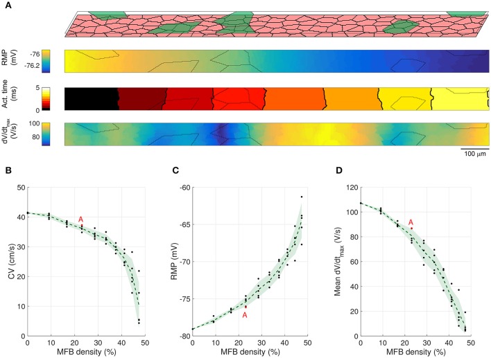
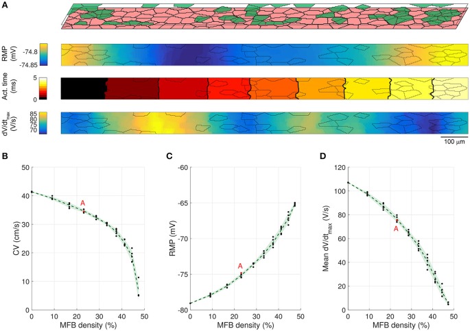
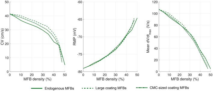
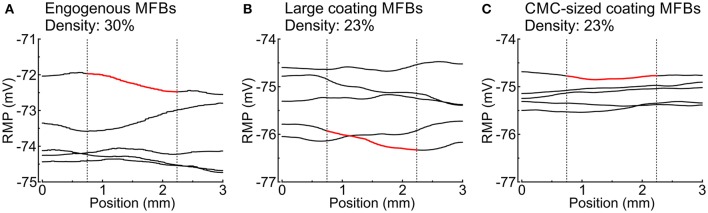
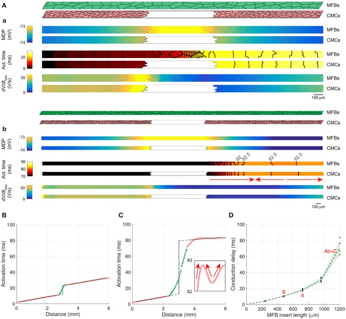
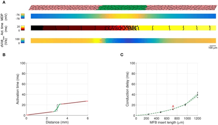
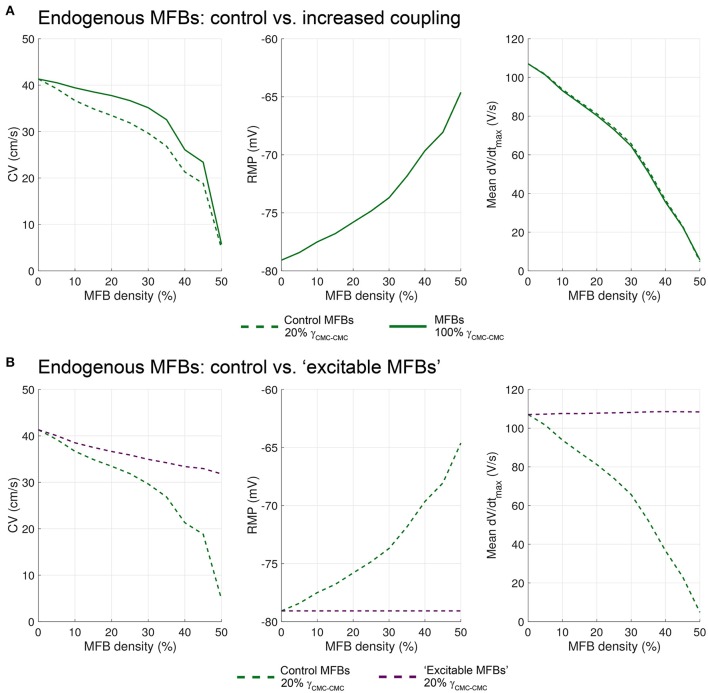
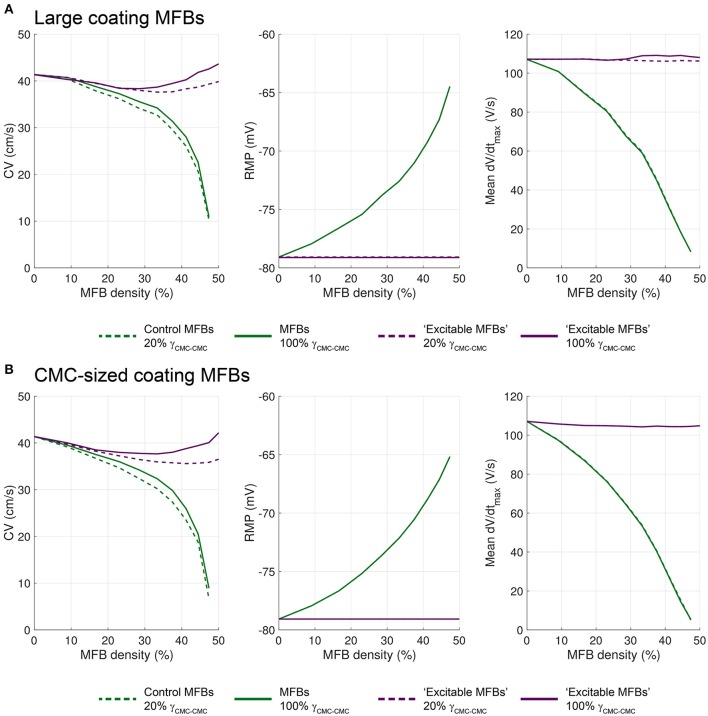
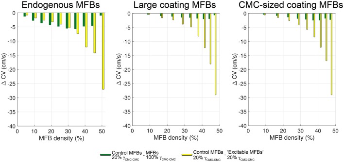
Fibrotic myocardial remodeling is typically accompanied by the appearance of myofibroblasts (MFBs). , MFBs were shown to slow conduction and precipitate ectopic activity following gap junctional coupling to cardiomyocytes (CMCs). To gain further mechanistic insights into this arrhythmogenic MFB-CMC crosstalk, we performed numerical simulations in cell-based high-resolution two-dimensional tissue models that replicated experimental conditions. Cell dimensions were determined using confocal microscopy of single and co-cultured neonatal rat ventricular CMCs and MFBs. Conduction was investigated as a function of MFB density in three distinct cellular tissue architectures: CMC strands with endogenous MFBs, CMC strands with coating MFBs of two different sizes, and CMC strands with MFB inserts. Simulations were performed to identify individual contributions of heterocellular gap junctional coupling and of the specific electrical phenotype of MFBs. With increasing MFB density, both endogenous and coating MFBs slowed conduction. At MFB densities of 5-30%, conduction slowing was most pronounced in strands with endogenous MFBs due to the MFB-dependent increase in axial resistance. At MFB densities >40%, very slow conduction and spontaneous activity was primarily due to MFB-induced CMC depolarization. Coating MFBs caused non-uniformities of resting membrane potential, which were more prominent with large than with small MFBs. In simulations of MFB inserts connecting two CMC strands, conduction delays increased with increasing insert lengths and block appeared for inserts >1.2 mm. Thus, electrophysiological properties of engineered CMC-MFB co-cultures depend on MFB density, MFB size and their specific positioning in respect to CMCs. These factors may influence conduction characteristics in the heterocellular myocardium.

摘要

纤维化心肌重塑通常伴随着肌成纤维细胞(MFBs)的出现。研究表明,MFBs与心肌细胞(CMCs)形成间隙连接偶联后会减慢传导并引发异位活动。为了进一步深入了解这种致心律失常的MFB-CMC串扰机制,我们在基于细胞的高分辨率二维组织模型中进行了数值模拟,该模型复制了实验条件。使用共聚焦显微镜对新生大鼠单个和共培养的心室CMCs和MFBs进行细胞尺寸测定。在三种不同的细胞组织结构中研究了传导与MFB密度的关系:具有内源性MFBs的CMC束、具有两种不同大小包被MFBs的CMC束以及具有MFB插入物的CMC束。进行模拟以确定异细胞间隙连接偶联和MFBs特定电表型的个体贡献。随着MFB密度的增加,内源性和包被MFBs均减慢传导。在MFB密度为5%-30%时,由于轴向电阻的MFB依赖性增加,具有内源性MFBs的束中传导减慢最为明显。在MFB密度>40%时,非常缓慢的传导和自发活动主要是由于MFB诱导的CMC去极化。包被MFBs导致静息膜电位不均匀,大MFBs比小MFBs更明显。在连接两条CMC束的MFB插入物模拟中,传导延迟随着插入物长度的增加而增加,对于长度>1.2 mm的插入物出现传导阻滞。因此,工程化CMC-MFB共培养物的电生理特性取决于MFB密度、MFB大小及其相对于CMCs的特定定位。这些因素可能会影响异细胞心肌中的传导特性。

https://cdn.ncbi.nlm.nih.gov/pmc/blobs/9933/5081362/4b977d7d7846/fphys-07-00496-g0013.jpg
https://cdn.ncbi.nlm.nih.gov/pmc/blobs/9933/5081362/a6820eb4a394/fphys-07-00496-g0001.jpg
https://cdn.ncbi.nlm.nih.gov/pmc/blobs/9933/5081362/a58e8127af64/fphys-07-00496-g0002.jpg
https://cdn.ncbi.nlm.nih.gov/pmc/blobs/9933/5081362/0659bd00d57f/fphys-07-00496-g0003.jpg
https://cdn.ncbi.nlm.nih.gov/pmc/blobs/9933/5081362/f7bc1084118d/fphys-07-00496-g0004.jpg
https://cdn.ncbi.nlm.nih.gov/pmc/blobs/9933/5081362/804da3134f8c/fphys-07-00496-g0005.jpg
https://cdn.ncbi.nlm.nih.gov/pmc/blobs/9933/5081362/2d7ef3d7e8fa/fphys-07-00496-g0006.jpg
https://cdn.ncbi.nlm.nih.gov/pmc/blobs/9933/5081362/d9679599421d/fphys-07-00496-g0007.jpg
https://cdn.ncbi.nlm.nih.gov/pmc/blobs/9933/5081362/e07fe441109d/fphys-07-00496-g0008.jpg
https://cdn.ncbi.nlm.nih.gov/pmc/blobs/9933/5081362/0fe1fa9a2983/fphys-07-00496-g0009.jpg
https://cdn.ncbi.nlm.nih.gov/pmc/blobs/9933/5081362/c3f102ef4e74/fphys-07-00496-g0010.jpg
https://cdn.ncbi.nlm.nih.gov/pmc/blobs/9933/5081362/204e2ae9f384/fphys-07-00496-g0011.jpg
https://cdn.ncbi.nlm.nih.gov/pmc/blobs/9933/5081362/bdabf0900627/fphys-07-00496-g0012.jpg
https://cdn.ncbi.nlm.nih.gov/pmc/blobs/9933/5081362/4b977d7d7846/fphys-07-00496-g0013.jpg

相似文献

1
Myofibroblasts Electrotonically Coupled to Cardiomyocytes Alter Conduction: Insights at the Cellular Level from a Detailed Tissue Structure Model.与心肌细胞电耦合的肌成纤维细胞改变传导:来自详细组织结构模型的细胞水平见解
Front Physiol. 2016 Oct 27;7:496. doi: 10.3389/fphys.2016.00496. eCollection 2016.
2
Aggravation of cardiac myofibroblast arrhythmogeneicity by mechanical stress.机械应力导致心肌成纤维细胞致心律失常性增强。
Cardiovasc Res. 2014 Dec 1;104(3):489-500. doi: 10.1093/cvr/cvu227. Epub 2014 Oct 24.
3
Connexin43 silencing in myofibroblasts prevents arrhythmias in myocardial cultures: role of maximal diastolic potential.缝隙连接蛋白 43 在肌成纤维细胞中的沉默可预防心肌培养中的心律失常:最大舒张电位的作用。
Cardiovasc Res. 2012 Mar 1;93(3):434-44. doi: 10.1093/cvr/cvr351. Epub 2011 Dec 22.
4
Effects of Na Current and Mechanogated Channels in Myofibroblasts on Myocyte Excitability and Repolarization.成肌纤维细胞中钠电流和机械门控通道对心肌细胞兴奋性和复极化的影响。
Comput Math Methods Med. 2016;2016:6189374. doi: 10.1155/2016/6189374. Epub 2016 Nov 17.
5
Susceptibility to arrhythmia in the infarcted heart depends on myofibroblast density.梗死心脏的心律失常易感性取决于成纤维细胞密度。
Biophys J. 2011 Sep 21;101(6):1307-15. doi: 10.1016/j.bpj.2011.08.009. Epub 2011 Sep 20.
6
A protective antiarrhythmic role of ursodeoxycholic acid in an in vitro rat model of the cholestatic fetal heart.熊去氧胆酸在胆汁淤积胎儿心脏体外大鼠模型中的保护抗心律失常作用。
Hepatology. 2011 Oct;54(4):1282-92. doi: 10.1002/hep.24492. Epub 2011 Aug 1.
7
Myofibroblast-Derived Exosomes Contribute to Development of a Susceptible Substrate for Atrial Fibrillation.肌成纤维细胞衍生的外泌体促成心房颤动易患基质的形成。
Cardiology. 2020;145(5):324-332. doi: 10.1159/000505641. Epub 2020 Apr 1.
8
Myofibroblasts in diseased hearts: new players in cardiac arrhythmias?患病心脏中的肌成纤维细胞:心律失常的新参与者?
Heart Rhythm. 2009 Jun;6(6):848-56. doi: 10.1016/j.hrthm.2009.02.038. Epub 2009 Feb 25.
9
Similar arrhythmicity in hypertrophic and fibrotic cardiac cultures caused by distinct substrate-specific mechanisms.不同基质特异性机制引起的肥厚和纤维化心脏培养中的相似心律失常性。
Cardiovasc Res. 2013 Jan 1;97(1):171-81. doi: 10.1093/cvr/cvs290. Epub 2012 Sep 12.
10
Abolishing myofibroblast arrhythmogeneicity by pharmacological ablation of α-smooth muscle actin containing stress fibers.通过药理学消融含有α-平滑肌肌动蛋白的应激纤维来消除肌成纤维细胞的致心律失常性。
Circ Res. 2011 Oct 28;109(10):1120-31. doi: 10.1161/CIRCRESAHA.111.244798. Epub 2011 Sep 15.

引用本文的文献

1
Computational Modeling of Effects of Gene Therapy on Ventricular Conduction Properties in Arrhythmogenic Cardiomyopathy.基因治疗对致心律失常性心肌病心室传导特性影响的计算模型
Circ Arrhythm Electrophysiol. 2025 May;18(5):e013658. doi: 10.1161/CIRCEP.124.013658. Epub 2025 Apr 9.
2
Myofibroblasts impair myocardial impulse propagation by heterocellular connexin43 gap-junctional coupling through micropores.肌成纤维细胞通过微孔的异细胞连接蛋白43间隙连接耦合损害心肌冲动传播。
Front Physiol. 2024 Feb 23;15:1352911. doi: 10.3389/fphys.2024.1352911. eCollection 2024.
3
Sacubitril/valsartan attenuates atrial conduction disturbance and electrophysiological heterogeneity with ameliorating fibrosis in mice.

本文引用的文献

1
Uncertainty and variability in computational and mathematical models of cardiac physiology.心脏生理学计算和数学模型中的不确定性与变异性。
J Physiol. 2016 Dec 1;594(23):6833-6847. doi: 10.1113/JP271671. Epub 2016 Jun 9.
2
Altered physiological functions and ion currents in atrial fibroblasts from patients with chronic atrial fibrillation.慢性心房颤动患者心房成纤维细胞的生理功能和离子电流改变。
Physiol Rep. 2016 Feb;4(2). doi: 10.14814/phy2.12681.
3
Simulation of Ectopic Pacemakers in the Heart: Multiple Ectopic Beats Generated by Reentry inside Fibrotic Regions.
沙库巴曲缬沙坦可减轻小鼠的心房传导障碍和电生理异质性,并改善纤维化。
Front Cardiovasc Med. 2024 Jan 19;11:1341601. doi: 10.3389/fcvm.2024.1341601. eCollection 2024.
4
Determinants of electrical propagation and propagation block in Arrhythmogenic Cardiomyopathy.致心律失常性心肌病中电传导和传导阻滞的决定因素。
J Mol Cell Cardiol. 2024 Jan;186:71-80. doi: 10.1016/j.yjmcc.2023.11.003. Epub 2023 Nov 11.
5
channelopathy: arrhythmia, cardiomyopathy, epilepsy and beyond.通道病:心律失常、心肌病、癫痫等。
Philos Trans R Soc Lond B Biol Sci. 2023 Jun 19;378(1879):20220164. doi: 10.1098/rstb.2022.0164. Epub 2023 May 1.
6
Editorial: Interplay between the heart and the immune system: Focus on heart rhythm regulation.社论:心脏与免疫系统之间的相互作用:关注心律调节。
Front Physiol. 2022 Aug 10;13:981499. doi: 10.3389/fphys.2022.981499. eCollection 2022.
7
Active force generation contributes to the complexity of spontaneous activity and to the response to stretch of murine cardiomyocyte cultures.主动力生成有助于自发性活动的复杂性,并有助于响应小鼠心肌细胞培养物的拉伸。
J Physiol. 2022 Jul;600(14):3287-3312. doi: 10.1113/JP283083. Epub 2022 Jun 23.
8
The Effects of Fibrotic Cell Type and Its Density on Atrial Fibrillation Dynamics: An In Silico Study.纤维母细胞类型及其密度对心房颤动动力学的影响:一项计算机模拟研究。
Cells. 2021 Oct 15;10(10):2769. doi: 10.3390/cells10102769.
9
Electroimmunology and cardiac arrhythmia.电免疫学与心律失常。
Nat Rev Cardiol. 2021 Aug;18(8):547-564. doi: 10.1038/s41569-021-00520-9. Epub 2021 Mar 2.
10
Optogenetic current in myofibroblasts acutely alters electrophysiology and conduction of co-cultured cardiomyocytes.肌成纤维细胞的光遗传学电流会急性改变共培养心肌细胞的电生理学和传导特性。
Sci Rep. 2021 Feb 24;11(1):4430. doi: 10.1038/s41598-021-83398-4.
心脏异位起搏点的模拟:纤维化区域内折返产生的多个异位搏动
Biomed Res Int. 2015;2015:713058. doi: 10.1155/2015/713058. Epub 2015 Oct 25.
4
Optical recording of calcium currents during impulse conduction in cardiac tissue.在心肌冲动传导过程中钙电流的光学记录。
Neurophotonics. 2015 Apr;2(2):021011. doi: 10.1117/1.NPh.2.2.021011. Epub 2015 Feb 13.
5
Aggravation of cardiac myofibroblast arrhythmogeneicity by mechanical stress.机械应力导致心肌成纤维细胞致心律失常性增强。
Cardiovasc Res. 2014 Dec 1;104(3):489-500. doi: 10.1093/cvr/cvu227. Epub 2014 Oct 24.
6
Nonlinear behaviour of conduction and block in cardiac tissue with heterogeneous expression of connexin 43.连接蛋白43表达不均一的心脏组织中传导和阻滞的非线性行为
J Mol Cell Cardiol. 2014 Nov;76:46-54. doi: 10.1016/j.yjmcc.2014.07.019. Epub 2014 Aug 12.
7
Spiral-wave dynamics in a mathematical model of human ventricular tissue with myocytes and fibroblasts.带有心肌细胞和成纤维细胞的人心室组织数学模型中的螺旋波动力学。
PLoS One. 2013 Sep 4;8(9):e72950. doi: 10.1371/journal.pone.0072950. eCollection 2013.
8
Gap junctions.间隙连接。
Compr Physiol. 2012 Jul;2(3):1981-2035. doi: 10.1002/cphy.c110051.
9
Microscopic variations in interstitial and intracellular structure modulate the distribution of conduction delays and block in cardiac tissue with source-load mismatch.间质和细胞内结构的微观变化调节了源-负载不匹配的心脏组织中传导延迟和阻滞的分布。
Europace. 2012 Nov;14 Suppl 5(Suppl 5):v3-v9. doi: 10.1093/europace/eus267.
10
A distinct de novo expression of Nav1.5 sodium channels in human atrial fibroblasts differentiated into myofibroblasts.在分化为肌成纤维细胞的人心房成纤维细胞中,Nav1.5 钠通道明显呈新表达。
J Physiol. 2012 Sep 1;590(17):4307-19. doi: 10.1113/jphysiol.2012.233593. Epub 2012 Jul 16.