Khadilkar Kranti, Sarathi Vijaya, Kasaliwal Rajeev, Pandit Reshma, Goroshi Manjunath, Malhotra Gaurav, Dalvi Abhay, Bakshi Ganesh, Bhansali Anil, Rajput Rajesh, Shivane Vyankatesh, Lila Anurag, Bandgar Tushar, Shah Nalini S
Department of EndocrinologySeth G S Medical College and KEM Hospital, Mumbai, India
Department of EndocrinologyVydehi Institute of Medical Sciences and Research Center, Bangalore, India.
Endocr Connect. 2016 Nov;5(6):89-97. doi: 10.1530/EC-16-0086. Epub 2016 Nov 16.
Malignant transformation of pheochromocytomas/paragangliomas (PCC/PGL) is a rare occurrence, and predictive factors for the same are not well understood. This study aims to identify the predictors of malignancy in patients with PCC/PGL.
We performed a retrospective analysis of 142 patients with either PCC or PGL registered at our institute between 2000 and 2015. Records were evaluated for clinical parameters like age, gender, familial/syndromic presentation, symptomatic presentation, biochemistry, size, number and location of tumours and presence of metastases and mode of its diagnosis.
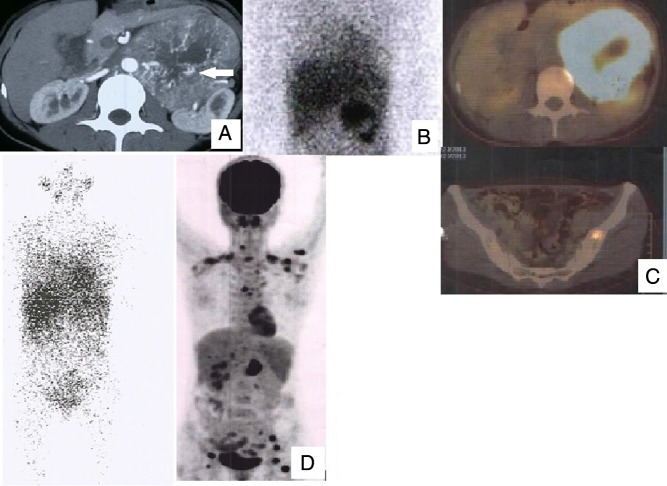
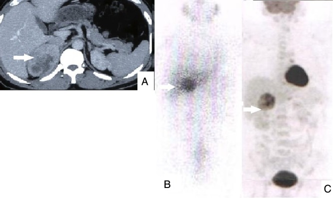
Twenty patients were found to have metastases; 13 had metastases at diagnosis and seven during follow-up. Metastases were detected by radiology (CT-neck to pelvis) in 11/20 patients (5/13 synchronous and 6/7 metachronous), I-metaiodobenzylguanidine in five (2/12 synchronous and 3/6 metachronous) patients and F-flurodeoxyglucose PET/CT in 15 (12/12 synchronous and 3/3 metachronous) patients. Malignant tumours were significantly larger than benign tumours (8.3 ± 4.1 cm, range: 3-22 cm vs 5.7 ± 2.3 cm, range: 2-14 cm, P = 0.0001) and less frequently metanephrine secreting. On linear regression analysis, tumour size and lack of metanephrine secretion were the independent predictors of malignancy.
Patients with primary tumour size >5.7 cm and lack of metanephrine secretory status should be evaluated for possible malignancy not only at diagnosis but also in the postoperative period. As compared to CT and I-MIBG scan, F-flurodeoxyglucose PET/CT analyses are better (sensitivity: 100%) for the diagnosis of metastases in our study.
嗜铬细胞瘤/副神经节瘤(PCC/PGL)的恶性转化较为罕见,其预测因素尚不明确。本研究旨在确定PCC/PGL患者恶性肿瘤的预测指标。
我们对2000年至2015年间在我院登记的142例PCC或PGL患者进行了回顾性分析。评估记录中的临床参数,如年龄、性别、家族性/综合征性表现、症状表现、生化指标、肿瘤大小、数量、位置以及转移情况及其诊断方式。
发现20例患者有转移;13例在诊断时已有转移,7例在随访期间出现转移。11/20例患者(5/13例为同步转移,6/7例为异时转移)通过放射学检查(颈部至骨盆CT)检测到转移,5例患者(2/12例为同步转移,3/6例为异时转移)通过间碘苄胍检测到转移,15例患者(12/12例为同步转移,3/3例为异时转移)通过氟脱氧葡萄糖PET/CT检测到转移。恶性肿瘤明显大于良性肿瘤(8.3±4.1cm,范围:3 - 22cm vs 5.7±2.3cm,范围:2 - 14cm,P = 0.0001),且分泌甲肾上腺素的频率较低。线性回归分析显示,肿瘤大小和缺乏甲肾上腺素分泌是恶性肿瘤的独立预测指标。
原发肿瘤大小>5.7cm且缺乏甲肾上腺素分泌状态的患者,不仅在诊断时,而且在术后均应评估是否可能存在恶性肿瘤。在我们的研究中,与CT和I - MIBG扫描相比,氟脱氧葡萄糖PET/CT分析对转移的诊断效果更好(敏感性:100%)。