Tang Jian-Cai, An Rui, Jiang Yi-Qing, Yang Jian
Department of Biochemistry, North of Sichuan Medical University, Nanchong, China.
School of Basic Medical Sciences, North of Sichuan Medical University, Nanchong, China.
Cancer Res Treat. 2017 Jul;49(3):778-789. doi: 10.4143/crt.2015.485. Epub 2016 Nov 11.
The purpose of this study was to observe the effects of metformin on human esophageal cancer cell and to investigate its possible mechanisms.
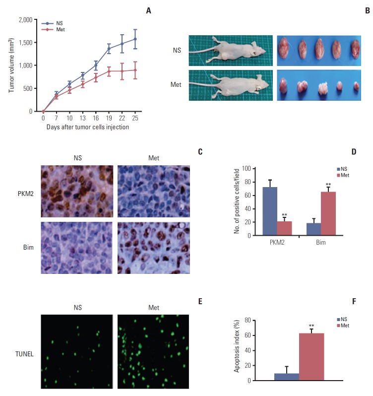
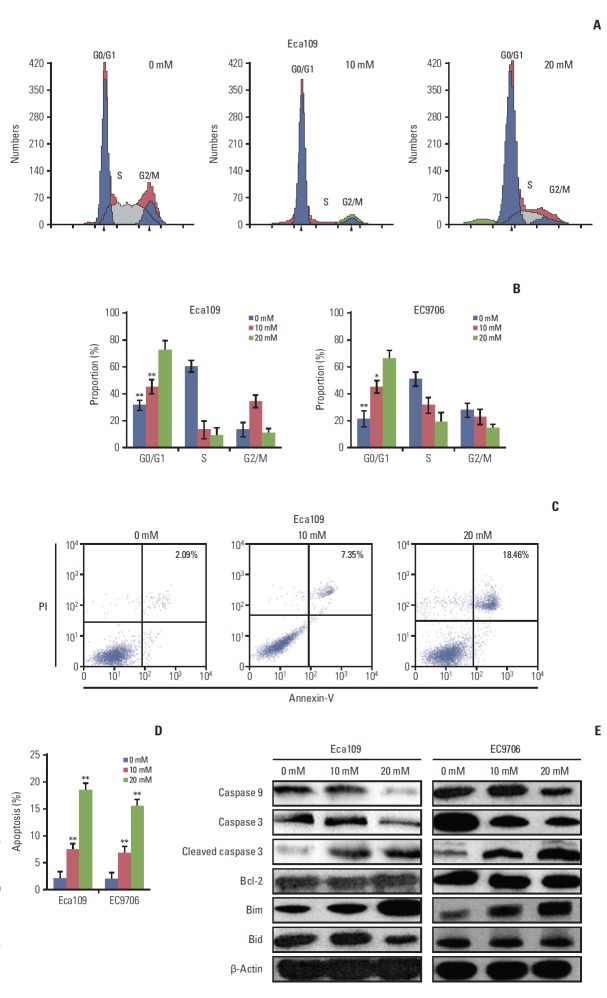
Cell viability was detected by using a Cell Counting Kit-8, while cell cycle and apoptosis were assessed by flow cytometry and western blot was used to measure the expression of the related proteins. RNAi was used to knockout pyruvate kinase muscle isozyme 2 (PKM2). An Eca109 tumor model was established to evaluate the antitumor effect . Immunohistochemistry was determined based on the expression of PKM2 and Bim in tumor tissues. Tunnel was used to assess tumor cell apoptosis.
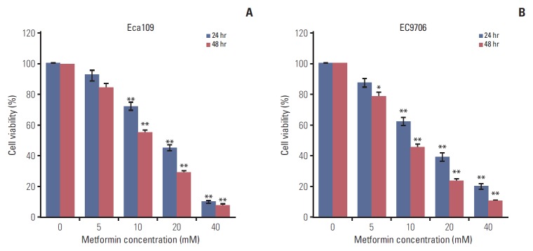
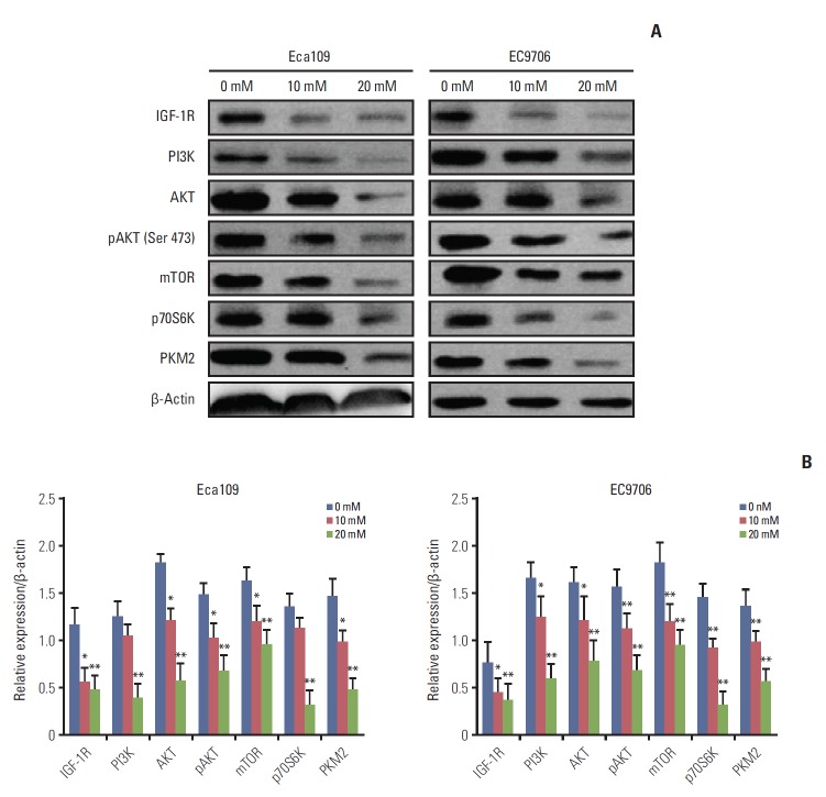
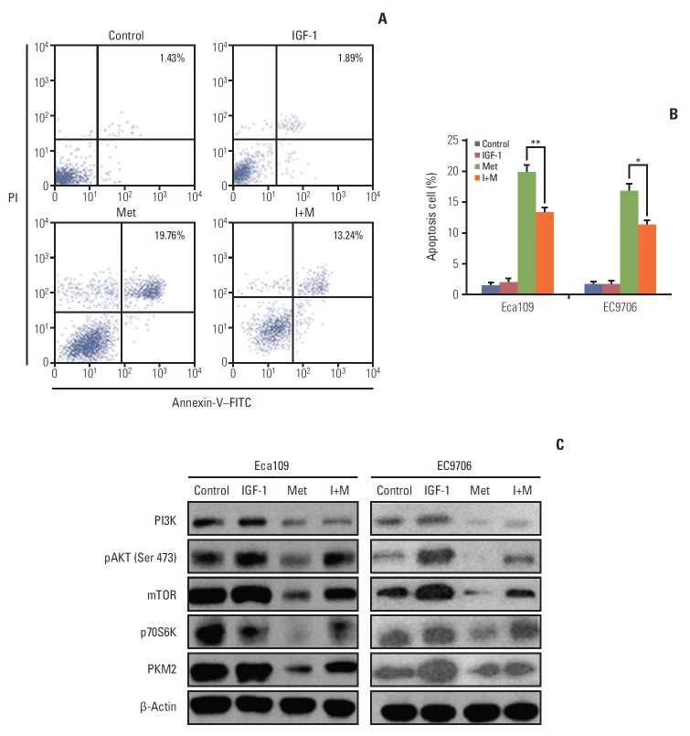
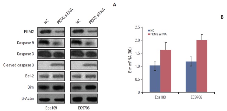
Esophageal cancer cells viability was reduced after metformin treatment. The cell cycle was arrested in the G0/G1 phase, apoptosis was induced, caspase 3 was activated, caspase 9 was downregulated, and the pro-apoptotic protein Bim increased. Further study revealed that metformin could suppress the expression of insulin-like growth factor 1 receptor and its downstream proteins, phosphoinositide 3-kinase (PI3K), protein kinase B (AKT/PKB), phosphorylation of AKT (pAKT), mammalian target of rapamycin (mTOR), p70S6K, and PKM2. Insulin-like growth factor 1 partly reversed metfromin-induced apoptosis and attenuated the repression effect of metfomin to PI3K, pAKT, and PKM2. Knockout PKM2 resulted in the activation of caspase 3, down-regulation of caspase 9, and increased expression of Bim. In the Eca109 xenograft model, metformin significantly reduced tumor growth. Furthermore, we found that metformin treatment increased the rate of apoptosis, down-regulation of PKM2, and up-regulation of Bim in tumor tissues.
Metformin restrained esophageal cancer cell proliferation partly by suppressing the PI3K/AKT/mTOR pathway.
本研究旨在观察二甲双胍对人食管癌细胞的作用,并探讨其可能的作用机制。
使用细胞计数试剂盒-8检测细胞活力,通过流式细胞术评估细胞周期和凋亡情况,采用蛋白质免疫印迹法检测相关蛋白的表达。利用RNA干扰技术敲除丙酮酸激酶M2型(PKM2)。建立Eca109肿瘤模型以评估抗肿瘤效果。基于肿瘤组织中PKM2和Bim的表达进行免疫组织化学检测。采用TUNEL法评估肿瘤细胞凋亡。
二甲双胍处理后食管癌细胞活力降低。细胞周期阻滞于G0/G1期,诱导凋亡,激活半胱天冬酶3,下调半胱天冬酶9,促凋亡蛋白Bim增加。进一步研究表明,二甲双胍可抑制胰岛素样生长因子1受体及其下游蛋白磷酸肌醇3激酶(PI3K)、蛋白激酶B(AKT/PKB)、磷酸化AKT(pAKT)、哺乳动物雷帕霉素靶蛋白(mTOR)、p70S6K和PKM2的表达。胰岛素样生长因子1部分逆转二甲双胍诱导的凋亡,并减弱二甲双胍对PI3K、pAKT和PKM2的抑制作用。敲除PKM2导致半胱天冬酶3激活,半胱天冬酶9下调,Bim表达增加。在Eca109异种移植模型中,二甲双胍显著抑制肿瘤生长。此外,我们发现二甲双胍处理可增加肿瘤组织中的凋亡率,下调PKM2,上调Bim。
二甲双胍部分通过抑制PI3K/AKT/mTOR信号通路抑制食管癌细胞增殖。