• 文献检索
  • 文档翻译
  • 深度研究
  • 学术资讯
  • Suppr Zotero 插件Zotero 插件
  • 邀请有礼
  • 套餐&价格
  • 历史记录
应用&插件
Suppr Zotero 插件Zotero 插件浏览器插件Mac 客户端Windows 客户端微信小程序
定价
高级版会员购买积分包购买API积分包
服务
文献检索文档翻译深度研究API 文档MCP 服务
关于我们
关于 Suppr公司介绍联系我们用户协议隐私条款
关注我们

Suppr 超能文献

核心技术专利:CN118964589B侵权必究
粤ICP备2023148730 号-1Suppr @ 2026

文献检索

告别复杂PubMed语法,用中文像聊天一样搜索,搜遍4000万医学文献。AI智能推荐,让科研检索更轻松。

立即免费搜索

文件翻译

保留排版,准确专业,支持PDF/Word/PPT等文件格式,支持 12+语言互译。

免费翻译文档

深度研究

AI帮你快速写综述,25分钟生成高质量综述,智能提取关键信息,辅助科研写作。

立即免费体验

小儿脊柱异常的超声检查与磁共振成像评估

Ultrasonography and magnetic resonance imaging evaluation of pediatric spinal anomalies.

作者信息

Dhingani Dhaval Durlabhbhai, Boruah Deb Kumar, Dutta Hemonta Kumar, Gogoi Rudra Kanta

机构信息

Department of Radio Diagnosis, Assam Medical College and Hospital, Dibrugarh, Assam, India.

Department of Pediatric Surgery, Assam Medical College and Hospital, Dibrugarh, Assam, India.

出版信息

J Pediatr Neurosci. 2016 Jul-Sep;11(3):206-212. doi: 10.4103/1817-1745.193374.

DOI:10.4103/1817-1745.193374
PMID:27857788
原文链接:https://pmc.ncbi.nlm.nih.gov/articles/PMC5108122/
Abstract

CONTEXT

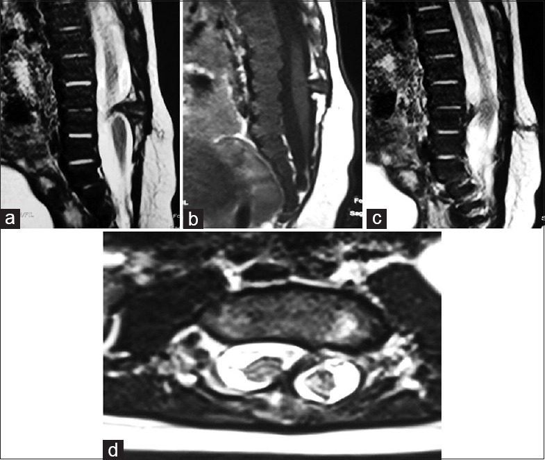
Spinal dysraphisms are congenital abnormalities of the spine due to imperfect fusion of midline mesenchymal, bony and neural structures. Imaging plays a vital role in their evaluation as significant portion of patients may present with concurrent anomalies that need to be corrected simultaneously to avoid repeat surgeries.

AIMS

The aims of the study were to evaluate Spinal dysraphisms using USG and MRI and to correlate imaging findings with operative findings in patients undergoing surgery.

SETTINGS AND DESIGN

Hospital based observational study conducted over a period of year.

MATERIALS AND METHODS

38 cases of both sexes and below 12 years of age with spinal dysraphism were studied. USG was performed in 29 cases where acoustic window was available for proper evaluation. MRI was performed in all cases. USG findings were compared with MRI findings and operative follow up was taken in 23 cases who underwent operative management.

STATISTICAL ANALYSIS USED

Results were analysed using percentage and arithmetic mean.

RESULTS

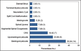
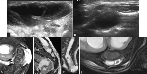
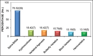
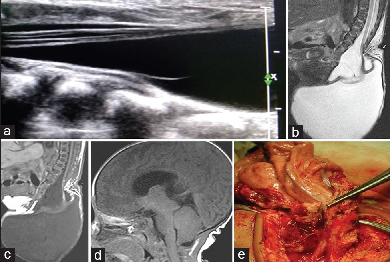
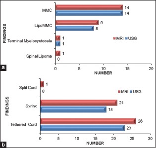
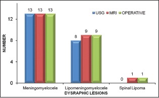
39.47 % cases were male and 60.53 % cases were female. Neonatal period was the most common presenting age group. Closed spinal dysraphism (63.16%) was more common than open (36.84%). 79.31% cases showed full agreement between spinal USG and MRI examinations and 6 out of 20.69% showed partial agreement. On operative correlation, USG findings were confirmatory in 91.30% cases and MRI findings were confirmatory in 100% cases.

CONCLUSIONS

USG can be used as the initial modality for evaluation of spinal dysraphism as well as for screening of suspected cases. MRI is indicated to confirm abnormal USG findings, which shows all concurrent abnormalities and also provides additional anatomical details relevant to surgical planning.

摘要

背景

脊柱裂是由于中线间充质、骨骼和神经结构融合不完全导致的先天性脊柱异常。影像学检查在其评估中起着至关重要的作用,因为相当一部分患者可能同时存在需要同时矫正的异常情况,以避免再次手术。

目的

本研究的目的是使用超声(USG)和磁共振成像(MRI)评估脊柱裂,并将影像学检查结果与接受手术治疗患者的手术结果进行关联。

设置与设计

基于医院的观察性研究,为期一年。

材料与方法

研究了38例年龄在12岁以下的脊柱裂患者,男女不限。29例有合适声学窗可进行恰当评估的患者进行了超声检查。所有病例均进行了MRI检查。将超声检查结果与MRI检查结果进行比较,并对23例接受手术治疗的患者进行了手术随访。

所用统计分析方法

结果采用百分比和算术平均值进行分析。

结果

男性患者占39.47%,女性患者占60.53%。新生儿期是最常见的发病年龄组。闭合性脊柱裂(63.16%)比开放性脊柱裂(36.84%)更常见。79.31%的病例脊柱超声检查和MRI检查结果完全一致,20.69%的病例中有6例部分一致。在手术相关性方面,超声检查结果在91.30%的病例中得到证实,MRI检查结果在100%的病例中得到证实。

结论

超声可作为评估脊柱裂以及筛查疑似病例的初始检查方法。对于超声检查异常结果,需行MRI检查以明确诊断,MRI可显示所有并发异常情况,并提供与手术规划相关的额外解剖细节。

https://cdn.ncbi.nlm.nih.gov/pmc/blobs/988d/5108122/8760cd1e5cff/JPN-11-206-g010.jpg
https://cdn.ncbi.nlm.nih.gov/pmc/blobs/988d/5108122/b5d930476c30/JPN-11-206-g001.jpg
https://cdn.ncbi.nlm.nih.gov/pmc/blobs/988d/5108122/558e8f0f361d/JPN-11-206-g002.jpg
https://cdn.ncbi.nlm.nih.gov/pmc/blobs/988d/5108122/c430809dfa9c/JPN-11-206-g003.jpg
https://cdn.ncbi.nlm.nih.gov/pmc/blobs/988d/5108122/8e1d064c4e1c/JPN-11-206-g004.jpg
https://cdn.ncbi.nlm.nih.gov/pmc/blobs/988d/5108122/0fe9d3b4434b/JPN-11-206-g005.jpg
https://cdn.ncbi.nlm.nih.gov/pmc/blobs/988d/5108122/2d2ed4e74e09/JPN-11-206-g007.jpg
https://cdn.ncbi.nlm.nih.gov/pmc/blobs/988d/5108122/1cee37e0a488/JPN-11-206-g008.jpg
https://cdn.ncbi.nlm.nih.gov/pmc/blobs/988d/5108122/d1d30e5a758a/JPN-11-206-g009.jpg
https://cdn.ncbi.nlm.nih.gov/pmc/blobs/988d/5108122/8760cd1e5cff/JPN-11-206-g010.jpg

相似文献

1
Ultrasonography and magnetic resonance imaging evaluation of pediatric spinal anomalies.小儿脊柱异常的超声检查与磁共振成像评估
J Pediatr Neurosci. 2016 Jul-Sep;11(3):206-212. doi: 10.4103/1817-1745.193374.
2
Spinal Dysraphisms: A New Anatomical-Clinicoradiological Classification.脊柱裂:一种新的解剖-临床放射学分类
Indian J Radiol Imaging. 2022 Jan 11;31(4):809-829. doi: 10.1055/s-0041-1741100. eCollection 2021 Oct.
3
Magnetic Resonance Imaging in Paediatric Spinal Dysraphism with Comparative Usefulness of Various Magnetic Resonance Sequences.小儿脊柱裂的磁共振成像及各种磁共振序列的比较效用
J Clin Diagn Res. 2017 Aug;11(8):TC17-TC22. doi: 10.7860/JCDR/2017/30134.10393. Epub 2017 Aug 1.
4
Differentiating Closed Versus Open Spinal Dysraphisms on Fetal MRI.胎儿磁共振成像中闭合性与开放性脊柱裂的鉴别诊断
AJR Am J Roentgenol. 2016 Dec;207(6):1316-1323. doi: 10.2214/AJR.16.16356. Epub 2016 Sep 9.
5
Spinal dysraphisms in the parturient: implications for perioperative anaesthetic care and labour analgesia.产妇的脊柱裂:对围手术期麻醉护理和分娩镇痛的影响。
Int J Obstet Anesth. 2015 Aug;24(3):252-63. doi: 10.1016/j.ijoa.2015.04.002. Epub 2015 Apr 18.
6
Unveiling the tale of the tail: an illustration of spinal dysraphisms.揭开尾巴的故事:脊柱发育异常的说明。
Neurosurg Rev. 2021 Feb;44(1):97-114. doi: 10.1007/s10143-019-01215-z. Epub 2019 Dec 7.
7
Comparison of ultrasound and magnetic resonance imaging findings in evaluation of fetal congenital anomalies: A single-institution prospective observational study.超声与磁共振成像在评估胎儿先天性异常中的表现比较:一项单机构前瞻性观察研究。
Med J Armed Forces India. 2023 Jul-Aug;79(4):439-450. doi: 10.1016/j.mjafi.2021.12.002. Epub 2022 Jan 21.
8
Unveiling the Integral Role of Magnetic Resonance Imaging in the Comprehensive Evaluation and Diagnosis of Spinal Dysraphism.揭示磁共振成像在脊柱裂综合评估与诊断中的重要作用。
Cureus. 2024 May 24;16(5):e60972. doi: 10.7759/cureus.60972. eCollection 2024 May.
9
Use of magnetic resonance imaging to detect occult spinal dysraphism in infants.利用磁共振成像检测婴儿隐匿性脊柱裂。
J Neurosurg Pediatr. 2017 Feb;19(2):217-226. doi: 10.3171/2016.8.PEDS16128. Epub 2016 Dec 2.
10
Prenatal and postnatal evaluation for syringomyelia in patients with spinal dysraphism.脊髓脊膜膨出患者脊髓空洞症的产前和产后评估。
J Neurosurg Pediatr. 2014 Sep;14(3):316-21. doi: 10.3171/2014.5.PEDS13482. Epub 2014 Jul 4.

引用本文的文献

1
Cloacal Dysgenesis Sequence in a Preterm Neonate.直肠生殖发育序列异常一例早产儿。
Am J Case Rep. 2024 Feb 27;25:e942203. doi: 10.12659/AJCR.942203.
2
MR Protocols for Paediatric Neurosurgical Common Conditions: An Update Guide for Neurosurgeons.儿科神经外科研讨会常见病例磁共振协议:神经外科医生更新指南。
Adv Tech Stand Neurosurg. 2023;48:57-72. doi: 10.1007/978-3-031-36785-4_3.
3
Occult spinal dysraphisms in newborns with skin markers: role of ultrasonography and magnetic resonance imaging.有皮肤标记的新生儿隐性脊柱闭合不全:超声检查和磁共振成像的作用

本文引用的文献

1
Lumbosacral dysraphism as cause of neurogenic bladder: Magnetic Resonance Imaging based study from SIUT Pakistan.腰骶部脊柱裂作为神经源性膀胱的病因:来自巴基斯坦 SIUT 的基于磁共振成像的研究
J Pak Med Assoc. 2015 May;65(5):501-5.
2
The accuracy of abnormal lumbar sonography findings in detecting occult spinal dysraphism: a comparison with magnetic resonance imaging.腰椎超声检查异常结果在检测隐性脊柱裂中的准确性:与磁共振成像的比较
J Neurosurg Pediatr. 2012 Aug;10(2):150-3. doi: 10.3171/2012.5.PEDS11564. Epub 2012 Jun 29.
3
Spinal dysraphism.脊柱裂
Childs Nerv Syst. 2018 Feb;34(2):285-291. doi: 10.1007/s00381-017-3638-0. Epub 2017 Oct 27.
4
Magnetic Resonance Imaging in Paediatric Spinal Dysraphism with Comparative Usefulness of Various Magnetic Resonance Sequences.小儿脊柱裂的磁共振成像及各种磁共振序列的比较效用
J Clin Diagn Res. 2017 Aug;11(8):TC17-TC22. doi: 10.7860/JCDR/2017/30134.10393. Epub 2017 Aug 1.
J Pediatr Neurosci. 2011 Oct;6(Suppl 1):S31-40. doi: 10.4103/1817-1745.85707.
4
Prospective study of spinal anomalies in children with infantile hemangiomas of the lumbosacral skin.腰骶部皮肤婴幼儿血管瘤患儿脊柱异常的前瞻性研究。
J Pediatr. 2010 Nov;157(5):789-94. doi: 10.1016/j.jpeds.2010.07.054. Epub 2010 Sep 9.
5
Spinal dysraphism with anorectal malformation: lumbosacral magnetic resonance imaging evaluation of 120 patients.脊柱发育不良合并肛门直肠畸形:120 例腰骶部磁共振成像评估。
J Pediatr Surg. 2010 Apr;45(4):769-76. doi: 10.1016/j.jpedsurg.2009.10.094.
6
Split cord malformations: a clinical study of 254 patients and a proposal for a new clinical-imaging classification.脊髓纵裂畸形:254例患者的临床研究及一种新的临床影像学分类建议
J Neurosurg. 2005 Dec;103(6 Suppl):531-6. doi: 10.3171/ped.2005.103.6.0531.
7
Spinal dysraphism: MR imaging rationale.脊柱裂:磁共振成像原理
J Neuroradiol. 2004 Jan;31(1):3-24. doi: 10.1016/s0150-9861(04)96875-7.
8
Multidisciplinary evaluation of occult spinal dysraphism in 47 children.47例儿童隐性脊柱裂的多学科评估
Scand J Urol Nephrol. 2003;37(4):329-34. doi: 10.1080/00365590310004725.
9
Spinal dysraphism: trends in northern India.脊柱裂:印度北部的趋势
Pediatr Neurosurg. 2003 Mar;38(3):133-45. doi: 10.1159/000068819.
10
Spinal ultrasound in infants.婴儿脊髓超声检查
Br J Radiol. 2002 Apr;75(892):384-92. doi: 10.1259/bjr.75.892.750384.