University of Washington, Seattle.
Arthritis Rheumatol. 2017 Apr;69(4):826-836. doi: 10.1002/art.39987.
Photosensitivity is common in patients with systemic lupus erythematosus, although the mechanisms linking ultraviolet (UV) light to flares are not well understood. We undertook this study to determine whether repetitive UVB exposure could induce type I interferon (IFN) production in normal mouse skin, and to investigate the roles of inflammatory monocytes and plasmacytoid dendritic cells (PDCs) in type I IFN production and development of UVB irradiation-induced inflammation.
Mice were irradiated with UVB at 100 mJ/cm for 5 days, and cutaneous manifestations were examined by messenger RNA expression of inflammatory and type I IFN response genes, histology, and flow cytometry. Inflammatory monocyte and PDC depletion experiments were performed in CCR2-diphtheria toxin receptor (DTR)-transgenic mice and blood dendritic cell antigen 2-DTR-transgenic mice. The roles of type I IFN and of the adaptor protein stimulator of IFN genes (STING) in UVB irradiation-induced inflammation were investigated using IFN-α/β/ω receptor (IFNAR)-knockout mice and STING-knockout mice.
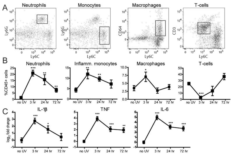
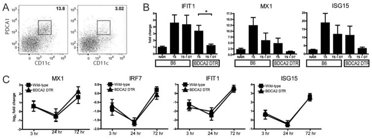
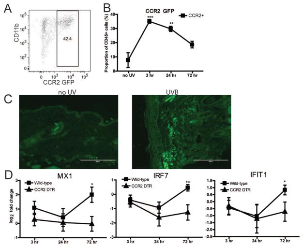
Repeated UVB irradiation stimulated an inflammatory cell infiltrate and induction of type I IFN and proinflammatory cytokines. Interestingly, the type I IFN response was independent of PDCs but dependent on inflammatory monocytes, which were recruited following UVB irradiation. The adaptor protein STING was necessary for both type I IFN and proinflammatory cytokine expression in the skin. UVB-irradiated IFNAR-knockout mice showed increased levels of proinflammatory genes and more severe inflammation by histology, suggesting a protective role for type I IFN.
In wild-type mice, repeated doses of UVB irradiation induce monocyte-dependent and PDC-independent expression of type I IFN together with expression of other proinflammatory cytokines. Induction is dependent on the adaptor protein STING. Surprisingly, studies using IFNAR-deficient mice revealed that type I IFN protects against UVB irradiation-induced skin inflammation, in part by attenuating proinflammatory cytokine expression and limiting tissue damage.
红斑狼疮患者常出现光过敏,尽管紫外线(UV)光与病情发作之间的关联机制尚未完全明确。我们开展此项研究旨在确定重复 UVB 照射是否会诱发正常小鼠皮肤产生 I 型干扰素(IFN),并探讨炎性单核细胞和浆细胞样树突状细胞(PDC)在 I 型 IFN 产生和 UVB 照射诱导炎症中的作用。
采用 100 mJ/cm2 的 UVB 照射小鼠 5 天,通过炎症和 I 型 IFN 反应基因的信使 RNA 表达、组织学和流式细胞术检测皮肤表现。在 CCR2-白喉毒素受体(DTR)转基因小鼠和血液树突状细胞抗原 2-DTR 转基因小鼠中进行炎性单核细胞和 PDC 耗竭实验。采用 IFN-α/β/ω 受体(IFNAR)敲除小鼠和 STING 敲除小鼠研究 I 型 IFN 和衔接蛋白干扰素基因刺激因子(STING)在 UVB 照射诱导炎症中的作用。
重复 UVB 照射可刺激炎症细胞浸润和 I 型 IFN 及促炎细胞因子的产生。有趣的是,I 型 IFN 反应不依赖于 PDC,但依赖于炎性单核细胞,后者在 UVB 照射后被募集。衔接蛋白 STING 对于皮肤中 I 型 IFN 和促炎细胞因子的表达都是必需的。UVB 照射的 IFNAR 敲除小鼠表现出促炎基因水平升高和组织学上更严重的炎症,表明 I 型 IFN 具有保护作用。
在野生型小鼠中,重复给予 UVB 照射可诱导单核细胞依赖性和 PDC 非依赖性的 I 型 IFN 表达,同时表达其他促炎细胞因子。诱导依赖于衔接蛋白 STING。令人惊讶的是,采用 IFNAR 缺陷型小鼠的研究表明,I 型 IFN 可防止 UVB 照射诱导的皮肤炎症,部分机制是通过减轻促炎细胞因子表达和限制组织损伤。