Kefayati Sarah, Amans Matthew, Faraji Farshid, Ballweber Megan, Kao Evan, Ahn Sinyeob, Meisel Karl, Halbach Van, Saloner David
Department of Radiology and Biomedical Imaging, UCSF, San Francisco, CA, USA.
Siemens Healthcare, CA, USA.
J Biomech. 2017 Jan 4;50:180-187. doi: 10.1016/j.jbiomech.2016.11.041. Epub 2016 Nov 15.
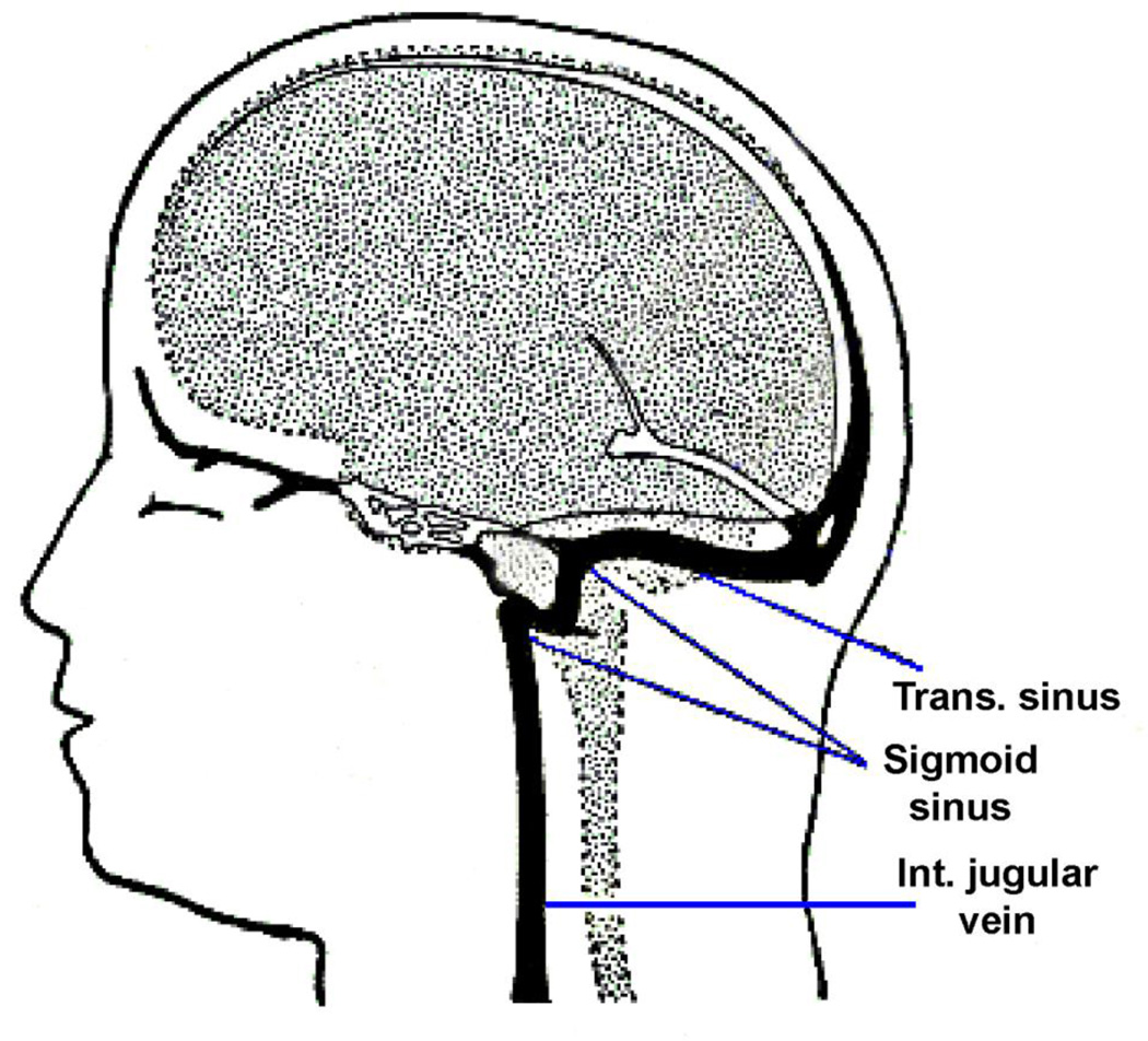
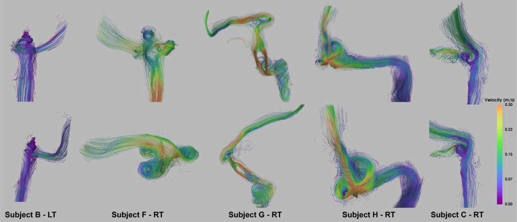
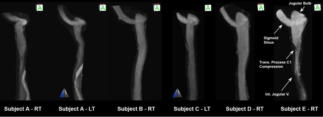
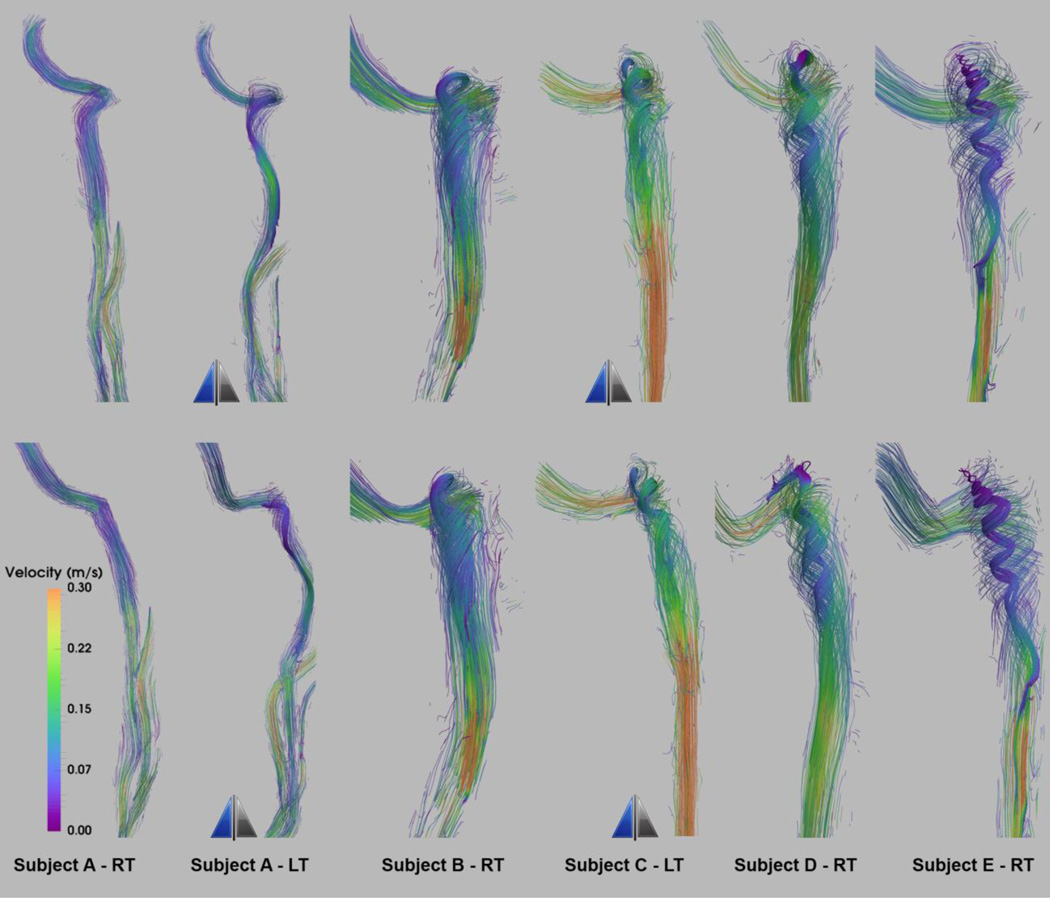
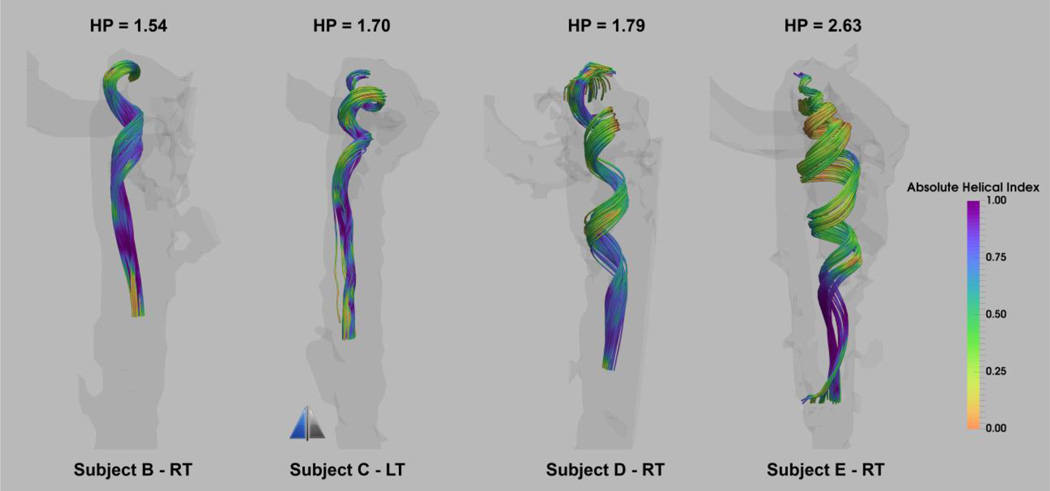
Aberrations in flow in the cerebral venous outflow tract (CVOT) have been implicated as the cause of several pathologic conditions including idiopathic intracranial hypertension (IIH), multiple sclerosis (MS), and pulsatile tinnitus (PT). The advent of 4D flow magnetic resonance imaging (4D-flow MRI) has recently allowed researchers to evaluate blood flow patterns in the arterial structures with great success. We utilized similar imaging techniques and found several distinct flow characteristics in the CVOT of subjects with and without lumenal irregularities. We present the flow patterns of 8 out of 38 subjects who have varying heights of the internal jugular bulb and varying lumenal irregularities including stenosis and diverticulum. In the internal jugular vein (IJV) with an elevated jugular bulb (JB), 4Dflow MRI revealed a characteristic spiral flow that was dependent on the level of JB elevation. Vortical flow was also observed in the diverticula of the venous sinuses and IJV. The diversity of flow complexity in the CVOT illustrates the potential importance of hemodynamic investigations in elucidating venous pathologies.
脑静脉流出道(CVOT)血流异常被认为是包括特发性颅内高压(IIH)、多发性硬化(MS)和搏动性耳鸣(PT)在内的多种病理状况的病因。4D流动磁共振成像(4D-flow MRI)的出现最近使研究人员能够非常成功地评估动脉结构中的血流模式。我们采用了类似的成像技术,在有和没有管腔不规则的受试者的CVOT中发现了几种不同的血流特征。我们展示了38名受试者中8名的血流模式,这些受试者的颈静脉球高度不同,管腔不规则情况各异,包括狭窄和憩室。在颈静脉球(JB)升高的颈内静脉(IJV)中,4D-flow MRI显示出一种特征性的螺旋血流,其取决于JB升高的程度。在静脉窦和IJV的憩室中也观察到了涡流。CVOT中血流复杂性的多样性说明了血流动力学研究在阐明静脉病变方面的潜在重要性。