Kao Evan, Kefayati Sarah, Amans Matthew R, Faraji Farshid, Ballweber Megan, Halbach Van, Saloner David
Department of Bioengineering, UC Berkeley, Berkeley, CA, USA; Department of Radiology and Biomedical Imaging, UCSF, San Francisco, CA, USA.
Department of Radiology and Biomedical Imaging, UCSF, San Francisco, CA, USA.
J Biomech. 2017 Feb 8;52:61-67. doi: 10.1016/j.jbiomech.2016.12.008. Epub 2016 Dec 14.
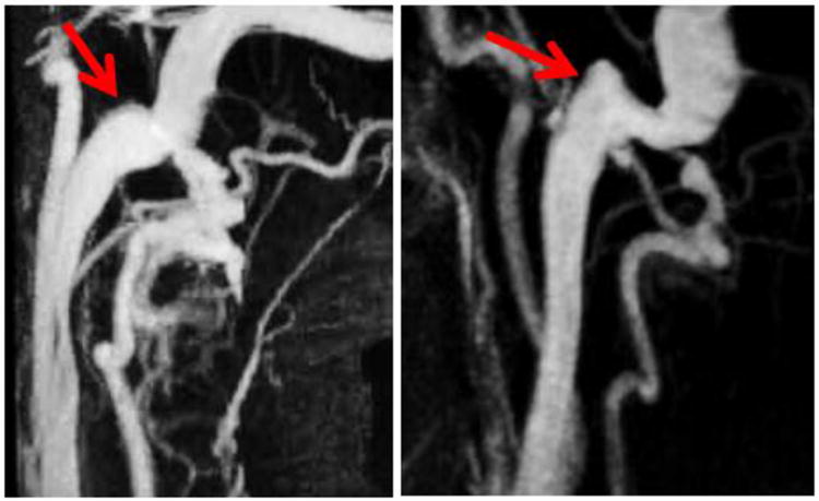
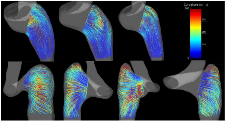
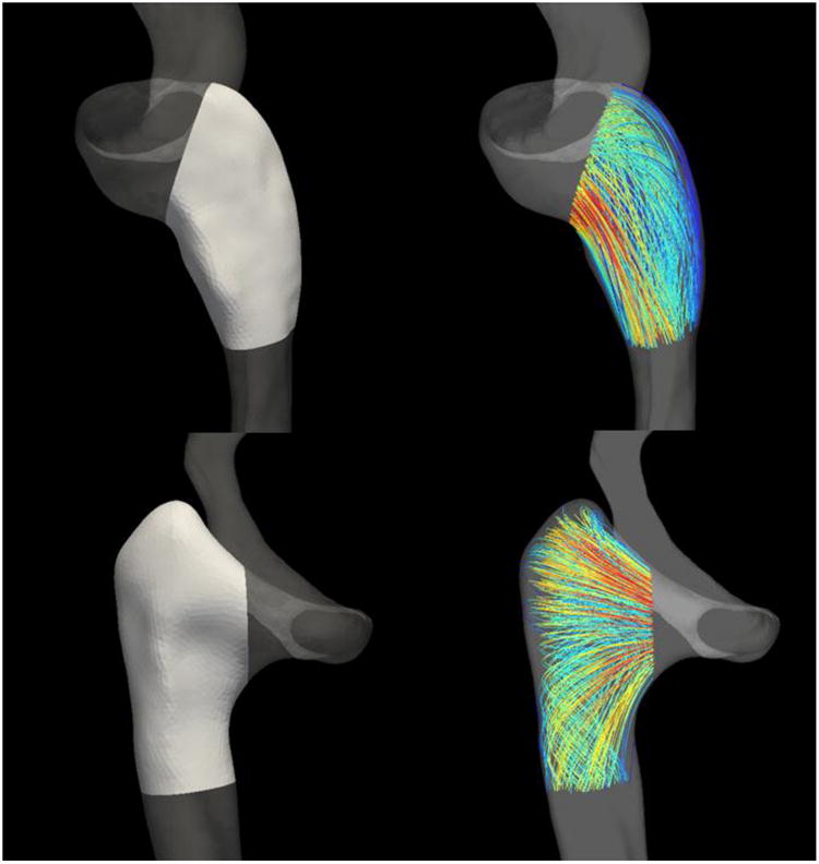
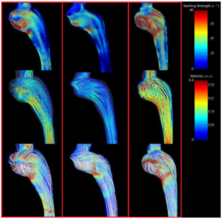
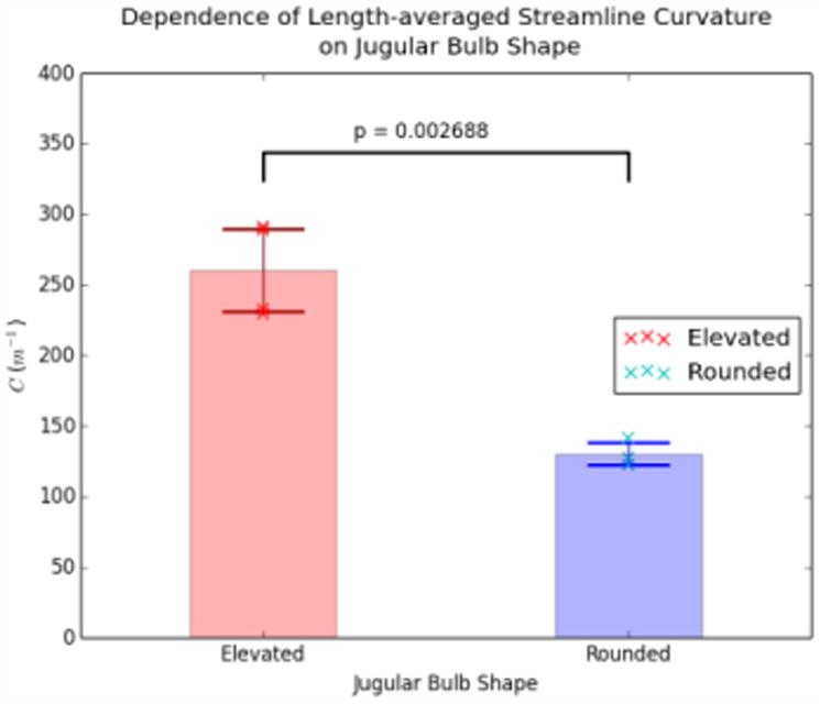
Pulsatile Tinnitus (PT) is a pulse-synchronous sound heard in the absence of an external source. PT is often related to abnormal flow in vascular structures near the cochlea. One vascular territory implicated in PT is the internal jugular vein (IJV). Using computational fluid dynamics (CFD) based on patient-specific Magnetic Resonance Imaging (MRI), we investigated the flow within the IJV of seven subjects, four symptomatic and three asymptomatic of PT. We found that there were two extreme anatomic types classified by the shape and position of the jugular bulbs: elevated and rounded. PT patients had elevated jugular bulbs that led to a distinctive helical flow pattern within the proximal internal jugular vein. Asymptomatic subjects generally had rounded jugular bulbs that neatly redirected flow from the sigmoid sinus directly into the jugular vein. These two flow patterns were quantified by calculating the length-averaged streamline curvature of the flow within the proximal jugular vein: 130.3±8.1m for geometries with rounded bulbs, 260.7±29.4m for those with elevated bulbs (P<0.005). Our results suggest that variations in the jugular bulb geometry lead to distinct flow patterns that are linked to PT, but further investigation is needed to determine if the vortex pattern is causal to sound generation.
搏动性耳鸣(PT)是一种在没有外部声源时听到的与脉搏同步的声音。PT通常与耳蜗附近血管结构中的异常血流有关。与PT相关的一个血管区域是颈内静脉(IJV)。我们基于患者特异性磁共振成像(MRI),使用计算流体动力学(CFD)研究了7名受试者颈内静脉内的血流情况,其中4名PT有症状患者,3名无症状患者。我们发现,根据颈静脉球的形状和位置可将其分为两种极端的解剖类型:高位型和圆形。PT患者的颈静脉球呈高位型,导致颈内静脉近端出现独特的螺旋血流模式。无症状受试者的颈静脉球通常为圆形,可将乙状窦的血流直接顺畅地导向颈静脉。通过计算颈静脉近端血流的长度平均流线曲率对这两种血流模式进行量化:圆形颈静脉球几何形状的为130.3±8.1m,高位颈静脉球几何形状的为260.7±29.4m(P<0.005)。我们的结果表明,颈静脉球几何形状的变化会导致与PT相关的不同血流模式,但需要进一步研究以确定涡流模式是否是产生声音的原因。