Cui Yanfen, Zhang Caiyuan, Luo Ran, Liu Huanhuan, Zhang Zhongyang, Xu Tianyong, Zhang Yong, Wang Dengbin
Department of Radiology, Xinhua Hospital, Shanghai Jiao Tong University School of Medicine.
MR Advanced Application and Research Center, GE Healthcare China, Shanghai, People's Republic of China.
Int J Nanomedicine. 2016 Nov 14;11:5671-5682. doi: 10.2147/IJN.S115357. eCollection 2016.
Arginine-glycine-aspartic acid (RGD)-based nanoprobes allow specific imaging of integrin αvβ3, a protein overexpressed during angiogenesis. Therefore, this study applied a novel RGD-coupled, polyacrylic acid (PAA)-coated ultrasmall superparamagnetic iron oxide (USPIO) (referred to as RGD-PAA-USPIO) in order to detect tumor angiogenesis and assess the early response to antiangiogenic treatment in human nasopharyngeal carcinoma (NPC) xenograft model by magnetic resonance imaging (MRI).
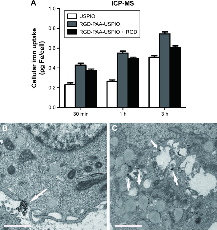
The binding specificity of RGD-PAA-USPIO with human umbilical vein endothelial cells (HUVECs) was confirmed by Prussian blue staining and transmission electron microscopy in vitro. The tumor targeting of RGD-PAA-USPIO was evaluated in the NPC xenograft model. Later, mice bearing NPC underwent MRI at baseline and after 4 and 14 days of consecutive treatment with Endostar or phosphate-buffered saline (n=10 per group).
The specific uptake of the RGD-PAA-USPIO nanoparticles was mainly dependent on the interaction between RGD and integrin αvβ3 of HUVECs. The tumor targeting of RGD-PAA-USPIO was observed in the NPC xenograft model. Moreover, the T2 relaxation time of mice in the Endostar-treated group decreased significantly compared with those in the control group both on days 4 and 14, consistent with the immunofluorescence results of CD31 and CD61 (<0.05).
This study demonstrated that the magnetic resonance molecular nanoprobes, RGD-PAA-USPIOs, allow noninvasive in vivo imaging of tumor angiogenesis and assessment of the early response to antiangiogenic treatment in NPC xenograft model, favoring its potential clinical translation.
基于精氨酸 - 甘氨酸 - 天冬氨酸(RGD)的纳米探针可对整合素αvβ3进行特异性成像,整合素αvβ3是血管生成过程中过度表达的一种蛋白质。因此,本研究应用了一种新型的RGD偶联、聚丙烯酸(PAA)包被的超小超顺磁性氧化铁(USPIO)(称为RGD - PAA - USPIO),以便通过磁共振成像(MRI)检测人鼻咽癌(NPC)异种移植模型中的肿瘤血管生成,并评估对抗血管生成治疗的早期反应。
通过普鲁士蓝染色和体外透射电子显微镜确认RGD - PAA - USPIO与人脐静脉内皮细胞(HUVECs)的结合特异性。在NPC异种移植模型中评估RGD - PAA - USPIO的肿瘤靶向性。随后,荷瘤小鼠在基线时以及连续用恩度或磷酸盐缓冲盐水治疗4天和14天后接受MRI检查(每组n = 10)。
RGD - PAA - USPIO纳米颗粒的特异性摄取主要取决于RGD与HUVECs整合素αvβ3之间的相互作用。在NPC异种移植模型中观察到RGD - PAA - USPIO的肿瘤靶向性。此外,恩度治疗组小鼠在第4天和第14天的T2弛豫时间均较对照组显著缩短,与CD31和CD61的免疫荧光结果一致(<0.05)。
本研究表明,磁共振分子纳米探针RGD - PAA - USPIO能够对NPC异种移植模型中的肿瘤血管生成进行无创体内成像,并评估对抗血管生成治疗的早期反应,有利于其潜在的临床转化。