Boord Peter, Madhyastha Tara M, Askren Mary K, Grabowski Thomas J
Department of Radiology, University of Washington, United States.
Department of Radiology, University of Washington, United States; Department of Neurology, University of Washington, United States.
Neuroimage Clin. 2016 Nov 5;13:1-8. doi: 10.1016/j.nicl.2016.11.004. eCollection 2017.
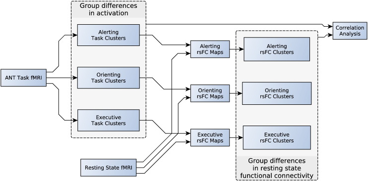
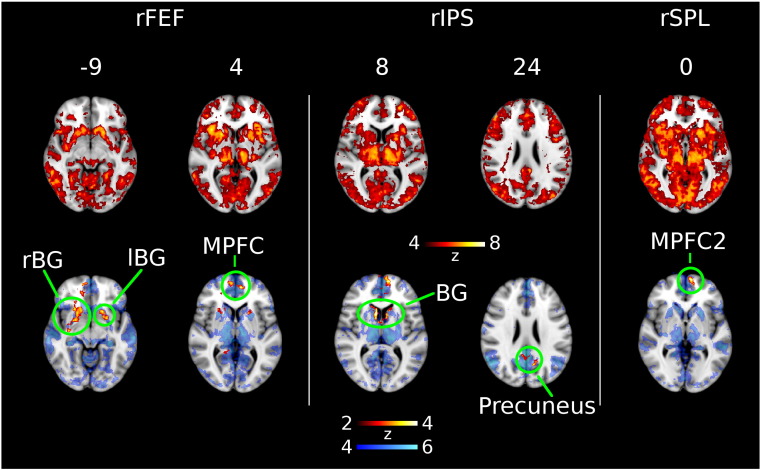
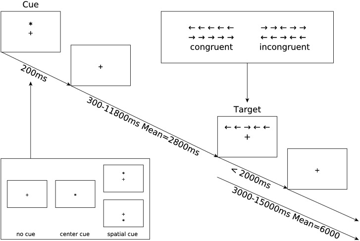
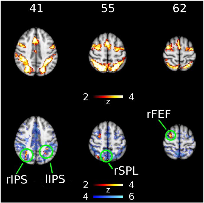
Attention dysfunction is a common but often undiagnosed cognitive impairment in Parkinson's disease that significantly reduces quality of life. We sought to increase understanding of the mechanisms underlying attention dysfunction using functional neuroimaging. Functional MRI was acquired at two repeated sessions in the resting state and during the Attention Network Test, for 25 non-demented subjects with Parkinson's disease and 21 healthy controls. Behavioral and MRI contrasts were calculated for alerting, orienting, and executive control components of attention. Brain regions showing group differences in attention processing were used as seeds in a functional connectivity analysis of a separate resting state run. Parkinson's disease subjects showed more activation during increased executive challenge in four regions of the dorsal attention and frontoparietal networks, namely right frontal eye field, left and right intraparietal sulcus, and precuneus. In three regions we saw reduced resting state connectivity to the default mode network. Further, whereas higher task activation in the right intraparietal sulcus correlated with reduced resting state connectivity between right intraparietal sulcus and the precuneus in healthy controls, this relationship was absent in Parkinson's disease subjects. Our results suggest that a weakened interaction between the default mode and task positive networks might alter the way in which the executive response is processed in PD.
注意力功能障碍是帕金森病中一种常见但往往未被诊断出的认知障碍,它会显著降低生活质量。我们试图通过功能神经影像学来加深对注意力功能障碍潜在机制的理解。对25名非痴呆帕金森病患者和21名健康对照者在静息状态和注意力网络测试期间的两个重复时段进行了功能磁共振成像(fMRI)检查。计算了注意力的警觉、定向和执行控制成分的行为和磁共振成像对比。在单独的静息状态扫描的功能连接分析中,将显示出注意力处理组间差异的脑区用作种子点。帕金森病患者在背侧注意力和额顶叶网络的四个区域(即右侧额叶眼区、左右顶内沟和楔前叶)执行挑战增加时表现出更多激活。在三个区域,我们观察到与默认模式网络的静息状态连接减少。此外,在健康对照者中,右侧顶内沟较高的任务激活与右侧顶内沟和楔前叶之间静息状态连接的减少相关,但在帕金森病患者中这种关系不存在。我们的结果表明,默认模式网络和任务正性网络之间减弱的相互作用可能会改变帕金森病中执行反应的处理方式。