Yang Zhiyong, Zhang Xinzhong, Guo Naipeng, Li Bin, Zhao Sheng
Department of Cardiology, Shengjing Hospital Affiliated to China Medical University, Shenyang, Liaoning, China (mainland).
Department of Cardiology, Capital Medical University Electric Power Teaching Hospital, Beijing, China (mainland).
Med Sci Monit. 2016 Dec 16;22:4937-4946. doi: 10.12659/msm.898454.
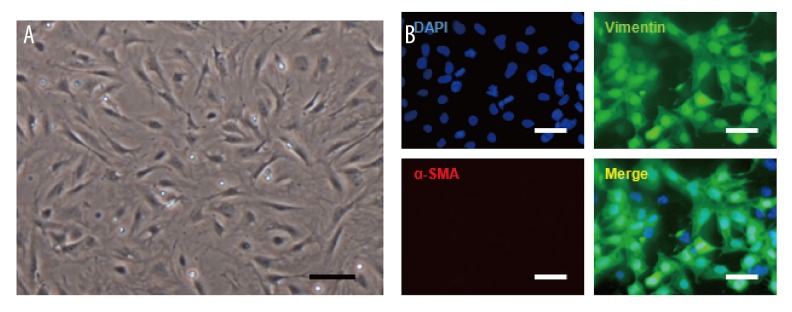
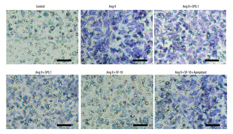
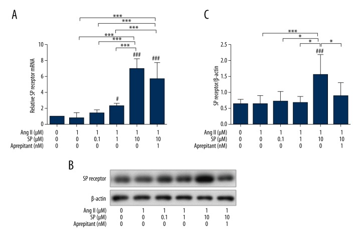
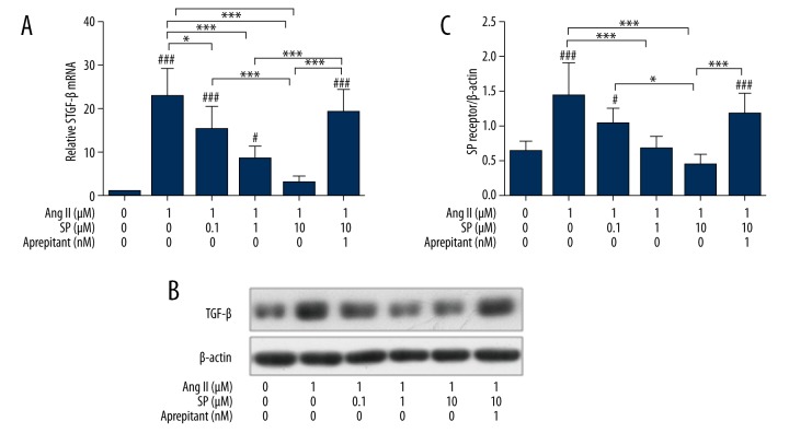
BACKGROUND The aim of this study was to explore the regulating effects of Substance P (SP) on the collagen synthesis of rat myocardial fibroblasts (CFBs) induced by angiotensin II (Ang II) and its potential mechanism. MATERIAL AND METHODS The CFBs of a neonatal SD rat were separately cultured and divided into the control group, Ang II treatment group, and treatment groups with different concentrations of SP, Ang II +; each group was given corresponding treatment respectively. RESULTS Ang II successfully induced the collagen synthesis of CFBs. Compared with the control group, the phosphorylation levels of TGF-β, erk, and smad2/3 were higher (p<0.05). Different concentrations of SP had an effect on Ang II-induced CFBs, reduced the collagen synthesis of CFBs, and increased the expressions of SP receptors, accompanied by lowering TGF-β protein, erk protein phosphorylation level, and smad2/3 protein phosphorylation level (p<0.05). Moreover, the higher the concentrations of SP, the more obvious of an effect it exerted. Treating the Ang II + SP group with aprepitant reduced the inhibiting effects of SP on collagen synthesis. The expression changes of collagen I and collagen III detected by immunocytochemistry were exactly in accordance with the results of qPCR and Western blotting. CONCLUSIONS SP can inhibit collagen synthesis of CFBs after Ang II inducing which may adjust the downstream signaling pathways associated protein including TGF-β, erk and smad2/3. SP can block the progress of myocardial fibrosis and is dose dependent, which is expected to be a promising target for the treatment of myocardial fibrosis.
背景 本研究旨在探讨P物质(SP)对血管紧张素II(Ang II)诱导的大鼠心肌成纤维细胞(CFB)胶原合成的调节作用及其潜在机制。
材料与方法 分离培养新生SD大鼠的CFB,分为对照组、Ang II处理组以及不同浓度SP处理组、Ang II +;每组分别给予相应处理。
结果 Ang II成功诱导CFB的胶原合成。与对照组相比,TGF-β、erk和smad2/3的磷酸化水平更高(p<0.05)。不同浓度的SP对Ang II诱导的CFB有影响,降低了CFB的胶原合成,并增加了SP受体的表达,同时降低了TGF-β蛋白、erk蛋白磷酸化水平和smad2/3蛋白磷酸化水平(p<0.05)。此外,SP浓度越高,作用越明显。用阿瑞匹坦处理Ang II + SP组可降低SP对胶原合成的抑制作用。免疫细胞化学检测的I型胶原和III型胶原表达变化与qPCR和蛋白质印迹结果完全一致。
结论 SP可抑制Ang II诱导后CFB的胶原合成,可能通过调节包括TGF-β、erk和smad2/3在内的下游信号通路相关蛋白发挥作用。SP可阻断心肌纤维化进程,且呈剂量依赖性,有望成为治疗心肌纤维化的有前景靶点。