Department of Integrative Medical Biology, Anatomy, Umeå University, SE-901 87, Umeå, Sweden.
Department of Community Medicine and Rehabilitation, Physiotherapy, Umeå University, Umeå, Sweden.
Stem Cell Res Ther. 2017 Nov 13;8(1):260. doi: 10.1186/s13287-017-0715-y.
We aimed to generate a bioengineered multi-lamellar human corneal stroma tissue in vitro by differentiating periodontal ligament stem cells (PDLSCs) towards keratocytes on an aligned silk membrane.
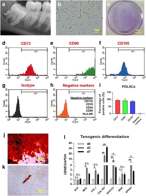
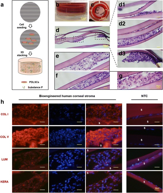
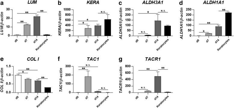
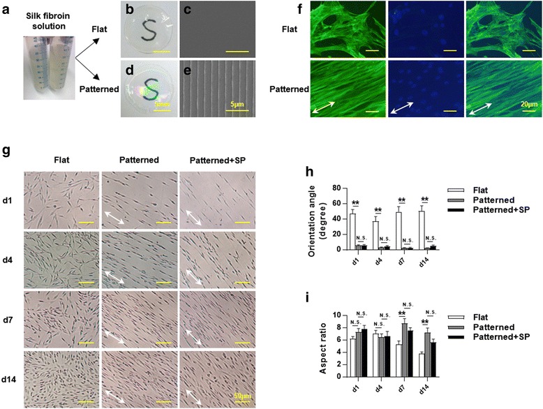
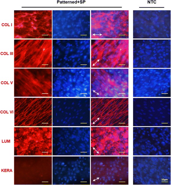
Human PDLSCs were isolated and identified. The neuropeptide substance P (SP) was added in keratocyte differentiation medium (KDM) to evaluate its effect on keratocyte differentiation of PDLSCs. PDLSCs were then seeded on patterned silk membrane and cultured with KDM and SP. Cell alignment was evaluated and the expression of extracellular matrix (ECM) components of corneal stroma was detected. Finally, multi-lamellar tissue was constructed in vitro by PDLSCs seeded on patterned silk membranes, which were stacked orthogonally and stimulated by KDM supplemented with SP for 18 days. Sections were prepared and subsequently stained with hematoxylin and eosin or antibodies for immunofluorescence observation of human corneal stroma-related proteins.
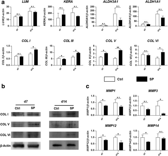
SP promoted the expression of corneal stroma-related collagens (collagen types I, III, V, and VI) during the differentiation induced by KDM. Patterned silk membrane guided cell alignment of PDLSCs, and important ECM components of the corneal stroma were shown to be deposited by the cells. The constructed multi-lamellar tissue was found to support cells growing between every two layers and expressing the main type of collagens (collagen types I and V) and proteoglycans (lumican and keratocan) of normal human corneal stroma.
Multi-lamellar human corneal stroma-like tissue can be constructed successfully in vitro by PDLSCs seeded on orthogonally aligned, multi-layered silk membranes with SP supplementation, which shows potential for future corneal tissue engineering.
我们旨在通过在取向丝膜上使牙周韧带干细胞(PDLSCs)向角膜细胞分化来体外生成生物工程化的多层人角膜基质组织。
分离和鉴定人牙周韧带干细胞。将神经肽物质 P(SP)添加到角膜细胞分化培养基(KDM)中,以评估其对 PDLSCs 向角膜细胞分化的影响。然后将 PDLSCs 接种在图案化丝膜上,并在 KDM 和 SP 存在的条件下培养。评估细胞取向,并检测角膜基质细胞外基质(ECM)成分的表达。最后,通过在 KDM 中补充 SP 刺激下将 PDLSCs 接种在图案化丝膜上构建体外多层组织,这些丝膜以正交方式堆叠并培养 18 天。制备切片并用苏木精和伊红或针对人角膜基质相关蛋白的免疫荧光观察的抗体进行染色。
SP 促进了 KDM 诱导的角膜基质相关胶原(I、III、V 和 VI 型胶原)的表达。图案化丝膜引导 PDLSCs 的细胞取向,并且细胞沉积了角膜基质的重要 ECM 成分。所构建的多层组织被发现支持细胞在每两层之间生长,并表达正常人类角膜基质的主要胶原(I 型和 V 型胶原)和蛋白聚糖(赖氨聚糖和角膜蛋白聚糖)类型。
通过在 SP 补充的正交定向、多层丝膜上接种 PDLSCs 可以成功在体外构建多层人角膜基质样组织,这为未来的角膜组织工程学提供了潜力。