Wang X, Lang M, Zhao T, Feng X, Zheng C, Huang C, Hao J, Dong J, Luo L, Li X, Lan C, Yu W, Yu M, Yang S, Ren H
Tianjin Medical University Cancer Institute and Hospital, National Clinical Research Center for Cancer; Key Laboratory of Cancer Prevention and Therapy, Department of Pancreatic Cancer, Tianjin, China.
The State Key Laboratory of Experimental Hematology, Institute of Hematology and Hospital of Blood Diseases, Chinese Academy of Medical Sciences and Peking Union Medical College, Tianjin, China.
Oncogene. 2017 May 25;36(21):3048-3058. doi: 10.1038/onc.2016.458. Epub 2016 Dec 19.
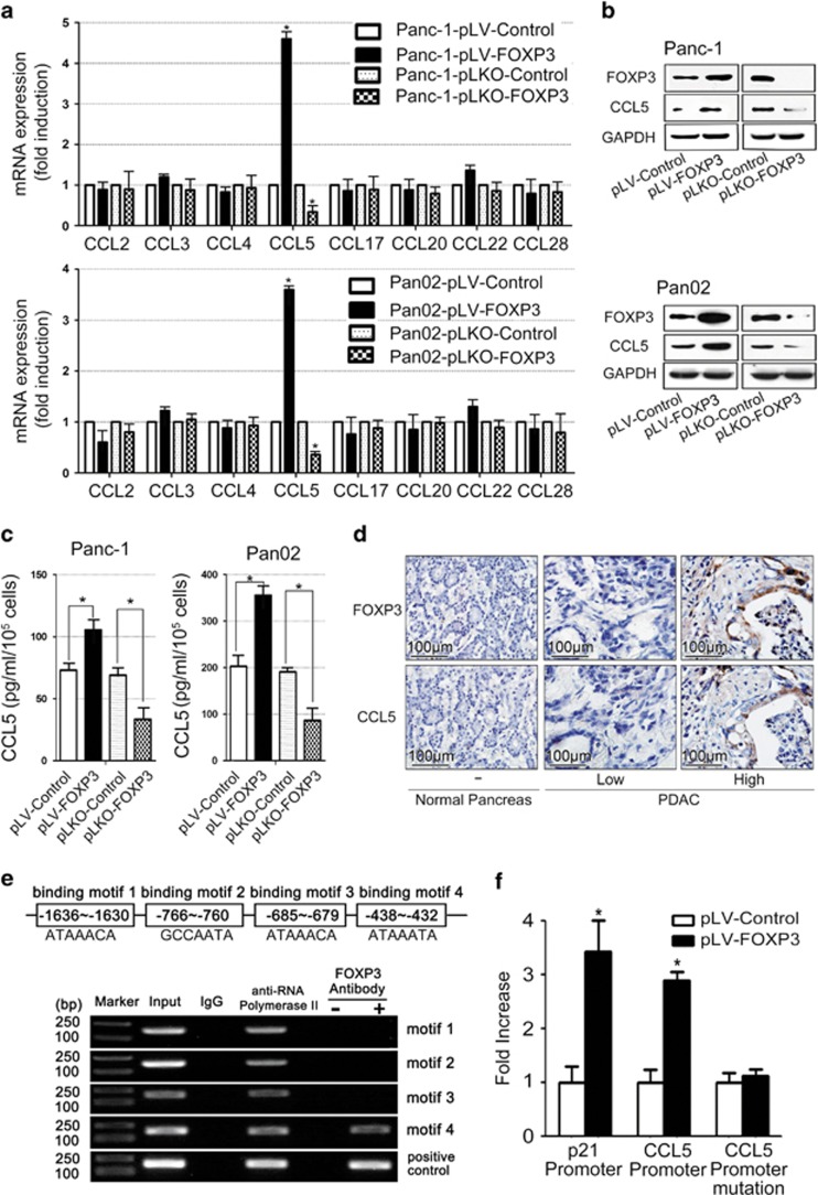
Forkheadbox protein 3 (FOXP3), initially identified as a key transcription factor for regulatory T cells (Treg cells), was also expressed in many tumors including pancreatic ductal adenocarcinoma (PDAC). However, its role in PDAC progression remains elusive. In this study, we utilized 120 PDAC tissues after radical resection to detect cancer-FOXP3 and Treg cells by immunohistochemistry and evaluated clinical and pathological features of these patients. Cancer-FOXP3 was positively correlated with Treg cells accumulation in tumor tissues derived from PDAC patients. In addition, high cancer-FOXP3 expression was associated with increased tumor volumes and poor prognosis in PDAC especially combined with high levels of Treg cells. Overexpression of cancer-FOXP3 promoted the tumor growth in immunocompetent syngeneic mice but not in immunocompromised or Treg cell-depleted mice. Furthermore, CCL5 was directly trans-activated by cancer-FOXP3 and promoted the recruitment of Treg cells from peripheral blood to the tumor site in vitro and in vivo. This finding has been further reinforced by the evidence that Treg cells recruitment by cancer-FOXP3 was impaired by neutralization of CCL5, thereby inhibiting the growth of PDAC. In conclusion, cancer-FOXP3 serves as a prognostic biomarker and a crucial determinant of immunosuppressive microenvironment via recruiting Treg cells by directly trans-activating CCL5. Therefore, cancer-FOXP3 could be used to select patients with better response to CCL5/CCR5 blockade immunotherapy.
叉头框蛋白3(FOXP3)最初被鉴定为调节性T细胞(Treg细胞)的关键转录因子,在包括胰腺导管腺癌(PDAC)在内的许多肿瘤中也有表达。然而,其在PDAC进展中的作用仍不清楚。在本研究中,我们利用120例根治性切除后的PDAC组织,通过免疫组织化学检测癌组织中的FOXP3和Treg细胞,并评估这些患者的临床和病理特征。癌组织中的FOXP3与PDAC患者肿瘤组织中Treg细胞的积累呈正相关。此外,癌组织中FOXP3高表达与PDAC肿瘤体积增大及预后不良相关,尤其是与高水平的Treg细胞同时存在时。癌组织中FOXP3的过表达促进了免疫健全的同基因小鼠的肿瘤生长,但在免疫缺陷或Treg细胞耗竭的小鼠中则不然。此外,CCL5可被癌组织中的FOXP3直接反式激活,并在体外和体内促进Treg细胞从外周血募集到肿瘤部位。CCL5的中和作用损害了癌组织中FOXP3介导的Treg细胞募集,从而抑制了PDAC的生长,这一证据进一步强化了这一发现。总之,癌组织中的FOXP3通过直接反式激活CCL5募集Treg细胞,作为一种预后生物标志物和免疫抑制微环境的关键决定因素。因此,癌组织中的FOXP3可用于选择对CCL5/CCR5阻断免疫疗法反应较好的患者。