• 文献检索
  • 文档翻译
  • 深度研究
  • 学术资讯
  • Suppr Zotero 插件Zotero 插件
  • 邀请有礼
  • 套餐&价格
  • 历史记录
应用&插件
Suppr Zotero 插件Zotero 插件浏览器插件Mac 客户端Windows 客户端微信小程序
定价
高级版会员购买积分包购买API积分包
服务
文献检索文档翻译深度研究API 文档MCP 服务
关于我们
关于 Suppr公司介绍联系我们用户协议隐私条款
关注我们

Suppr 超能文献

核心技术专利:CN118964589B侵权必究
粤ICP备2023148730 号-1Suppr @ 2026

文献检索

告别复杂PubMed语法,用中文像聊天一样搜索,搜遍4000万医学文献。AI智能推荐,让科研检索更轻松。

立即免费搜索

文件翻译

保留排版,准确专业,支持PDF/Word/PPT等文件格式,支持 12+语言互译。

免费翻译文档

深度研究

AI帮你快速写综述,25分钟生成高质量综述,智能提取关键信息,辅助科研写作。

立即免费体验

血管性轻度认知障碍和遗忘型轻度认知障碍之间的白质微观结构有何不同?

How does white matter microstructure differ between the vascular and amnestic mild cognitive impairment?

作者信息

Yu Yang, Liang Xinyu, Yu Haikuo, Zhao Weina, Lu Yan, Huang Yue, Yin Changhao, Gong Gaolang, Han Ying

机构信息

Department of Neurology, Hongqi Hospital of Mudanjiang Medical University, Mudanjiang, Heilongjiang, China.

State Key Laboratory of Cognitive Neuroscience and Learning & IDG/McGovern Institute for Brain Research, Beijing Normal University, Beijing, China.

出版信息

Oncotarget. 2017 Jan 3;8(1):42-50. doi: 10.18632/oncotarget.13960.

DOI:10.18632/oncotarget.13960
PMID:27992372
原文链接:https://pmc.ncbi.nlm.nih.gov/articles/PMC5352131/
Abstract

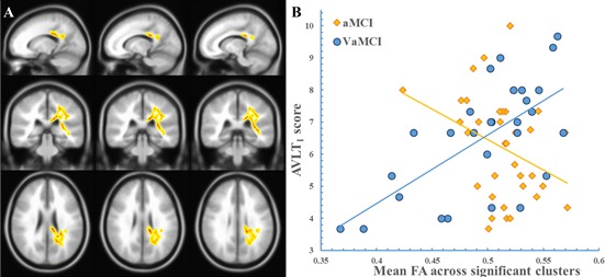
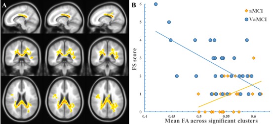
Changes in white matter (WM) microstructure may relate to the pathophysiology of cognitive impairment. Whether WM microstructure differs in two common pre-dementia subtypes, vascular mild cognitive impairment (VaMCI) and amnestic mild cognitive impairment (aMCI), is largely unknown. This study included 28 VaMCI (12 men, age: 46 ~ 77 years) and 34 aMCI patients (14 men, age: 51 ~ 79 years). All patients underwent a battery of neuropsychological tests and structural and diffusion magnetic resonance imaging (MRI) scanning. WM microstructure was quantified using diffusion MRI parameters: fractional anisotropy (FA), mean diffusivity (MD), axial diffusivity (AxD) and radial diffusivity (RD). These parameters were compared between the two patient groups using tract-based spatial statistics (TBSS) after controlling for age, gender, and education. No significant differences in FA/MD/AxD/RD were observed between the VaMCI and aMCI groups, which suggests a similar pattern of WM microstructure in the early stage of cognitive impairment for different dementia types. However, the two groups exhibited significant differences in the relationship between FA and the Auditory Verbal Learning Test (AVLT), which were primarily located around the corona radiate and corpus callosum. Specifically, there were significant positive correlations (R = 0.64, P < 0.001) between the FA and AVLT in the VaMCI group, but the opposite trend was observed in the aMCI group (R = -0.34, P = 0.047). The differential relationship between WM and memory between VaMCI and aMCI indicates an independent neuropathology for specific memory deficits in different types of dementia.

摘要

白质(WM)微观结构的变化可能与认知障碍的病理生理学有关。在两种常见的痴呆前期亚型,即血管性轻度认知障碍(VaMCI)和遗忘型轻度认知障碍(aMCI)中,WM微观结构是否存在差异在很大程度上尚不清楚。本研究纳入了28例VaMCI患者(12例男性,年龄:4677岁)和34例aMCI患者(14例男性,年龄:5179岁)。所有患者均接受了一系列神经心理学测试以及结构和扩散磁共振成像(MRI)扫描。使用扩散MRI参数对白质微观结构进行量化:各向异性分数(FA)、平均扩散率(MD)、轴向扩散率(AxD)和径向扩散率(RD)。在控制年龄、性别和教育程度后,使用基于体素的空间统计学(TBSS)对两组患者的这些参数进行比较。在VaMCI组和aMCI组之间未观察到FA/MD/AxD/RD的显著差异,这表明不同痴呆类型在认知障碍早期的WM微观结构模式相似。然而,两组在FA与听觉言语学习测试(AVLT)之间的关系上存在显著差异,主要位于放射冠和胼胝体周围。具体而言,VaMCI组中FA与AVLT之间存在显著正相关(R = 0.64,P < 0.001),但在aMCI组中观察到相反的趋势(R = -0.34,P = 0.047)。VaMCI和aMCI之间WM与记忆的差异关系表明不同类型痴呆中特定记忆缺陷存在独立的神经病理学机制。

https://cdn.ncbi.nlm.nih.gov/pmc/blobs/c602/5352131/0f3b6e042200/oncotarget-08-42-g002.jpg
https://cdn.ncbi.nlm.nih.gov/pmc/blobs/c602/5352131/4a20b47ec2d9/oncotarget-08-42-g001.jpg
https://cdn.ncbi.nlm.nih.gov/pmc/blobs/c602/5352131/0f3b6e042200/oncotarget-08-42-g002.jpg
https://cdn.ncbi.nlm.nih.gov/pmc/blobs/c602/5352131/4a20b47ec2d9/oncotarget-08-42-g001.jpg
https://cdn.ncbi.nlm.nih.gov/pmc/blobs/c602/5352131/0f3b6e042200/oncotarget-08-42-g002.jpg

相似文献

1
How does white matter microstructure differ between the vascular and amnestic mild cognitive impairment?血管性轻度认知障碍和遗忘型轻度认知障碍之间的白质微观结构有何不同?
Oncotarget. 2017 Jan 3;8(1):42-50. doi: 10.18632/oncotarget.13960.
2
White matter degeneration in subjective cognitive decline: a diffusion tensor imaging study.主观认知衰退中的白质退变:一项扩散张量成像研究
Oncotarget. 2016 Aug 23;7(34):54405-54414. doi: 10.18632/oncotarget.10091.
3
Amnestic mild cognitive impairment in Parkinson's disease: White matter structural changes and mechanisms.帕金森病遗忘型轻度认知障碍:白质结构变化及其机制。
PLoS One. 2019 Dec 12;14(12):e0226175. doi: 10.1371/journal.pone.0226175. eCollection 2019.
4
White matter integrity disruption in the pre-dementia stages of Alzheimer's disease: from subjective memory impairment to amnestic mild cognitive impairment.阿尔茨海默病痴呆前阶段的脑白质完整性破坏:从主观记忆损害到遗忘型轻度认知障碍。
Eur J Neurol. 2019 May;26(5):800-807. doi: 10.1111/ene.13892. Epub 2019 Jan 23.
5
Cortical Thickness and Microstructural White Matter Changes Detect Amnestic Mild Cognitive Impairment.皮质厚度和微观结构白质变化可检测遗忘型轻度认知障碍。
J Alzheimers Dis. 2017;56(1):415-428. doi: 10.3233/JAD-160724.
6
Genetic variation in angiotensin converting-enzyme affects the white matter integrity and cognitive function of amnestic mild cognitive impairment patients.血管紧张素转换酶基因多态性影响遗忘型轻度认知障碍患者的脑白质完整性和认知功能。
J Neurol Sci. 2017 Sep 15;380:177-181. doi: 10.1016/j.jns.2017.06.026. Epub 2017 Jun 16.
7
Identification of Amnestic Mild Cognitive Impairment Using Multi-Modal Brain Features: A Combined Structural MRI and Diffusion Tensor Imaging Study.利用多模态脑特征识别遗忘型轻度认知障碍:一项结构磁共振成像与扩散张量成像联合研究
J Alzheimers Dis. 2015;47(2):509-22. doi: 10.3233/JAD-150184.
8
[Changes in Plasma Amyloid-β Level and Their Relationship With White Matter Microstructure in Patients With Mild Cognitive Impairment].[轻度认知障碍患者血浆淀粉样蛋白β水平的变化及其与白质微结构的关系]
Zhongguo Yi Xue Ke Xue Yuan Xue Bao. 2023 Aug;45(4):571-580. doi: 10.3881/j.issn.1000-503X.15418.
9
[Study on the white matter neuronal integrity in amnestic mild cognitive impairment based on automating fiber-tract quantification].基于自动纤维束定量分析的遗忘型轻度认知障碍患者白质神经元完整性研究
Zhonghua Yi Xue Za Zhi. 2020 Jan 21;100(3):172-177. doi: 10.3760/cma.j.issn.0376-2491.2020.03.003.
10
White Matter Integrity in Subcortical Vascular Cognitive Impairment: A Multimodal Structural MRI Study.皮质下血管性认知障碍中的白质完整性:一项多模态结构磁共振成像研究
Curr Alzheimer Res. 2017;14(9):991-999. doi: 10.2174/1567205014666170329113707.

引用本文的文献

1
Multimodality neuroimaging in vascular mild cognitive impairment: A narrative review of current evidence.血管性轻度认知障碍的多模态神经影像学:当前证据的叙述性综述
Front Aging Neurosci. 2023 Mar 15;15:1073039. doi: 10.3389/fnagi.2023.1073039. eCollection 2023.
2
The clinical profile of cerebral small vessel disease: Toward an evidence-based identification of cognitive markers.脑小血管病的临床特征:基于证据的认知标志物识别。
Alzheimers Dement. 2023 Jan;19(1):244-260. doi: 10.1002/alz.12650. Epub 2022 Mar 31.
3
Distinctive Alterations of Functional Connectivity Strength between Vascular and Amnestic Mild Cognitive Impairment.

本文引用的文献

1
Brain structural, functional, and cognitive correlates of recent versus remote autobiographical memories in amnestic Mild Cognitive Impairment.遗忘型轻度认知障碍中近期与远期自传体记忆的脑结构、功能及认知相关性
Neuroimage Clin. 2015 May 28;8:473-82. doi: 10.1016/j.nicl.2015.05.010. eCollection 2015.
2
The Impact of Memory Change on Daily Life in Normal Aging and Mild Cognitive Impairment.正常衰老和轻度认知障碍中记忆变化对日常生活的影响。
Gerontologist. 2016 Oct;56(5):877-85. doi: 10.1093/geront/gnv030. Epub 2015 Apr 9.
3
Microstructural White Matter Abnormalities and Cognitive Dysfunction in Subcortical Ischemic Vascular Disease: an Atlas-Based Diffusion Tensor Analysis Study.
血管性和遗忘型轻度认知障碍之间功能连接强度的显著改变。
Neural Plast. 2021 May 19;2021:8812490. doi: 10.1155/2021/8812490. eCollection 2021.
4
Changes in Gray Matter Asymmetries of the Fusiform and Parahippocampal Gyruses in Patients With Subcortical Ischemic Vascular Disease.皮质下缺血性血管疾病患者梭状回和海马旁回灰质不对称性的变化
Front Neurol. 2021 Jan 21;11:603977. doi: 10.3389/fneur.2020.603977. eCollection 2020.
5
Disrupted Brain Structural Connectivity Network in Subcortical Ischemic Vascular Cognitive Impairment With No Dementia.无痴呆的皮质下缺血性血管性认知障碍中脑结构连接网络受损
Front Aging Neurosci. 2020 Jan 29;12:6. doi: 10.3389/fnagi.2020.00006. eCollection 2020.
6
Progressively Disrupted Brain Functional Connectivity Network in Subcortical Ischemic Vascular Cognitive Impairment Patients.皮质下缺血性血管性认知障碍患者大脑功能连接网络的渐进性破坏
Front Neurol. 2018 Feb 26;9:94. doi: 10.3389/fneur.2018.00094. eCollection 2018.
皮质下缺血性血管疾病中的微观结构白质异常与认知功能障碍:一项基于图谱的扩散张量分析研究
J Mol Neurosci. 2015 Jun;56(2):363-70. doi: 10.1007/s12031-015-0550-5. Epub 2015 Apr 10.
4
Diagnostic criteria for vascular cognitive disorders: a VASCOG statement.血管性认知障碍的诊断标准:VASCOG声明
Alzheimer Dis Assoc Disord. 2014 Jul-Sep;28(3):206-18. doi: 10.1097/WAD.0000000000000034.
5
White matter hyperintensities in mild cognitive impairment: clinical impact of location and interaction with lacunes and medial temporal atrophy.轻度认知障碍中的脑白质高信号:位置的临床影响及其与腔隙性脑梗死和海马萎缩的相互作用。
J Stroke Cerebrovasc Dis. 2014 May-Jun;23(5):e365-72. doi: 10.1016/j.jstrokecerebrovasdis.2013.12.040. Epub 2014 Feb 28.
6
The influence of cerebral small vessel disease on default mode network deactivation in mild cognitive impairment.脑小血管病对轻度认知障碍默认模式网络去激活的影响。
Neuroimage Clin. 2012 Nov 16;2:33-42. doi: 10.1016/j.nicl.2012.11.005. eCollection 2012.
7
Diffusion tensor imaging measures of normal appearing white matter in patients who are aging, or have amnestic mild cognitive impairment, or Alzheimer's disease.正常老年人、遗忘型轻度认知障碍患者和阿尔茨海默病患者的脑白质各向异性弥散张量成像测量。
J Clin Neurosci. 2013 Aug;20(8):1089-94. doi: 10.1016/j.jocn.2012.09.025. Epub 2013 Jun 17.
8
White matter changes in patients with amnestic mild cognitive impairment detected by diffusion tensor imaging.利用弥散张量成像技术检测遗忘型轻度认知障碍患者的脑白质改变。
PLoS One. 2013;8(3):e59440. doi: 10.1371/journal.pone.0059440. Epub 2013 Mar 21.
9
PANDA: a pipeline toolbox for analyzing brain diffusion images.PANDA:一个用于分析大脑扩散图像的管道工具包。
Front Hum Neurosci. 2013 Feb 21;7:42. doi: 10.3389/fnhum.2013.00042. eCollection 2013.
10
Structural and functional changes in subcortical vascular mild cognitive impairment: a combined voxel-based morphometry and resting-state fMRI study.皮质下血管性轻度认知障碍的结构和功能变化:一项基于体素形态计量学和静息态 fMRI 的联合研究。
PLoS One. 2012;7(9):e44758. doi: 10.1371/journal.pone.0044758. Epub 2012 Sep 19.