Li Yinghua, Guo Min, Lin Zhengfang, Zhao Mingqi, Xiao Misi, Wang Changbing, Xu Tiantian, Chen Tianfeng, Zhu Bing
Center Laboratory, Guangzhou Women and Children's Medical Center, Guangzhou Medical University.
Department of Chemistry, Jinan University, Guangzhou, People's Republic of China.
Int J Nanomedicine. 2016 Dec 8;11:6693-6702. doi: 10.2147/IJN.S122666. eCollection 2016.
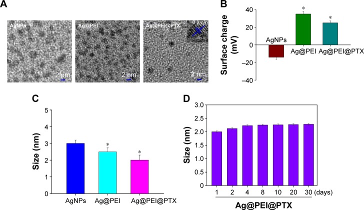
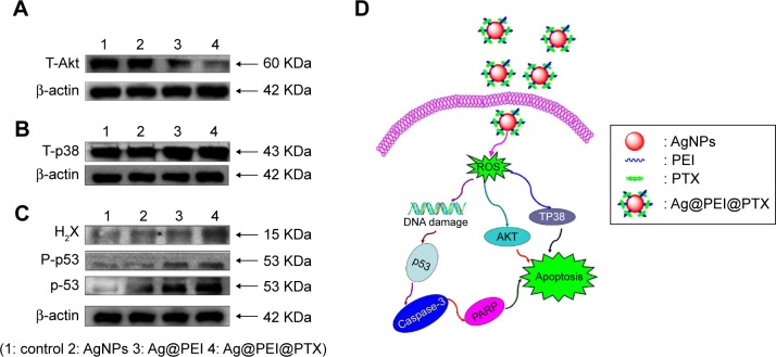
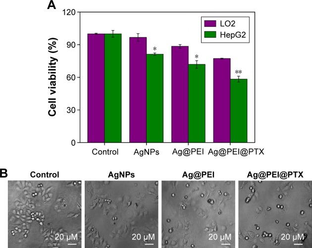
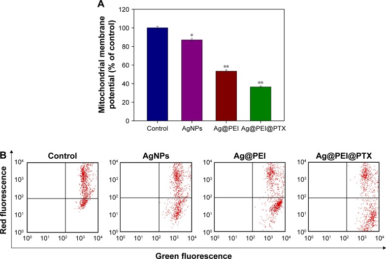
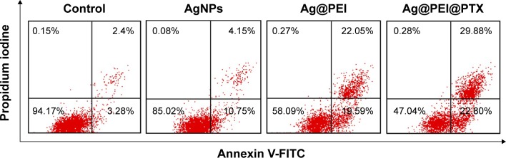
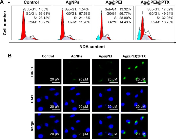
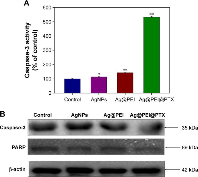
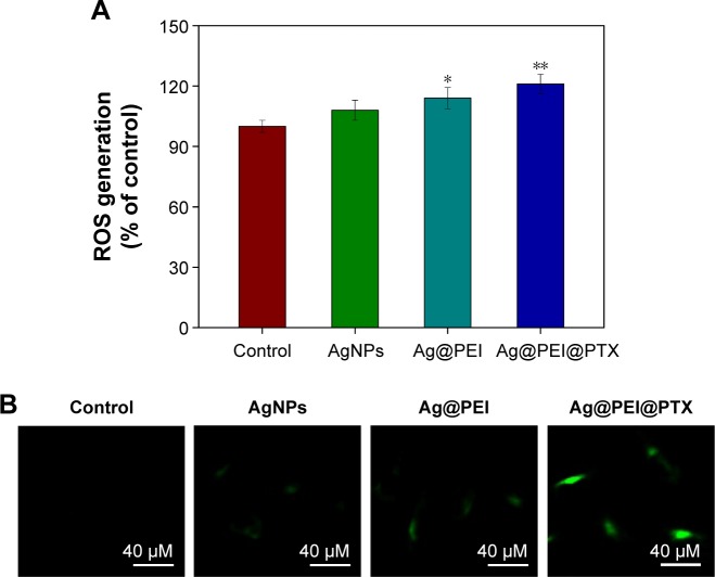
Hepatocarcinoma is the third leading cause of cancer-related deaths around the world. Recently, a novel emerging nanosystem as anticancer therapeutic agents with intrinsic therapeutic properties has been widely used in various medical applications. In this study, surface decoration of functionalized silver nanoparticles (AgNPs) by polyethylenimine (PEI) and paclitaxel (PTX) was synthesized. The purpose of this study was to evaluate the effect of Ag@ PEI@PTX on cytotoxic and anticancer mechanism on HepG2 cells. The transmission electron microscope image and 3-(4,5-dimethylthiazol-2-yl)-2,5-diphenyltetrazolium bromide assay showed that Ag@PEI@PTX had satisfactory size distribution and high stability and selectivity between cancer and normal cells. Ag@PEI@PTX-induced HepG2 cell apoptosis was confirmed by accumulation of the sub-G1 cells population, translocation of phosphatidylserine, depletion of mitochondrial membrane potential, DNA fragmentation, caspase-3 activation, and poly(ADP-ribose) polymerase cleavage. Furthermore, Ag@PEI@PTX enhanced cytotoxic effects on HepG2 cells and triggered intracellular reactive oxygen species; the signaling pathways of AKT, p53, and MAPK were activated to advance cell apoptosis. In conclusion, the results reveal that Ag@ PEI@PTX may provide useful information on Ag@PEI@PTX-induced HepG2 cell apoptosis and as appropriate candidate for chemotherapy of cancer.
肝癌是全球癌症相关死亡的第三大主要原因。最近,一种新型的具有内在治疗特性的纳米系统作为抗癌治疗剂已被广泛应用于各种医学领域。在本研究中,通过聚乙烯亚胺(PEI)和紫杉醇(PTX)对功能化银纳米颗粒(AgNPs)进行表面修饰并合成。本研究的目的是评估Ag@PEI@PTX对HepG2细胞的细胞毒性和抗癌机制的影响。透射电子显微镜图像和3-(4,5-二甲基噻唑-2-基)-2,5-二苯基四氮唑溴盐检测表明,Ag@PEI@PTX具有令人满意的尺寸分布,在癌细胞和正常细胞之间具有高稳定性和选择性。通过亚G1期细胞群体的积累、磷脂酰丝氨酸的易位、线粒体膜电位的耗竭、DNA片段化、半胱天冬酶-3激活和聚(ADP-核糖)聚合酶裂解,证实了Ag@PEI@PTX诱导的HepG2细胞凋亡。此外,Ag@PEI@PTX增强了对HepG2细胞的细胞毒性作用并引发细胞内活性氧;激活了AKT、p53和MAPK信号通路以促进细胞凋亡。总之,结果表明Ag@PEI@PTX可能为Ag@PEI@PTX诱导的HepG2细胞凋亡提供有用信息,并作为癌症化疗的合适候选物。