• 文献检索
  • 文档翻译
  • 深度研究
  • 学术资讯
  • Suppr Zotero 插件Zotero 插件
  • 邀请有礼
  • 套餐&价格
  • 历史记录
应用&插件
Suppr Zotero 插件Zotero 插件浏览器插件Mac 客户端Windows 客户端微信小程序
定价
高级版会员购买积分包购买API积分包
服务
文献检索文档翻译深度研究API 文档MCP 服务
关于我们
关于 Suppr公司介绍联系我们用户协议隐私条款
关注我们

Suppr 超能文献

核心技术专利:CN118964589B侵权必究
粤ICP备2023148730 号-1Suppr @ 2026

文献检索

告别复杂PubMed语法,用中文像聊天一样搜索,搜遍4000万医学文献。AI智能推荐,让科研检索更轻松。

立即免费搜索

文件翻译

保留排版,准确专业,支持PDF/Word/PPT等文件格式,支持 12+语言互译。

免费翻译文档

深度研究

AI帮你快速写综述,25分钟生成高质量综述,智能提取关键信息,辅助科研写作。

立即免费体验

大鼠胶质瘤模型中联合抗血管生成与放射治疗后MRI追踪的肿瘤血管参数的急性时间变化:确定协同作用的特征

Acute Temporal Changes of MRI-Tracked Tumor Vascular Parameters after Combined Anti-angiogenic and Radiation Treatments in a Rat Glioma Model: Identifying Signatures of Synergism.

作者信息

Elmghirbi Rasha, Nagaraja Tavarekere N, Brown Stephen L, Panda Swayamprava, Aryal Madhava P, Keenan Kelly A, Bagher-Ebadian Hassan, Cabral Glauber, Ewing James R

机构信息

a   Department of Physics, Oakland University, Rochester, Michigan.

b   Department of Neurology, enry Ford Hospital, Detroit, Michigan.

出版信息

Radiat Res. 2017 Jan;187(1):79-88. doi: 10.1667/RR14358.1. Epub 2016 Dec 21.

DOI:10.1667/RR14358.1
PMID:28001908
原文链接:https://pmc.ncbi.nlm.nih.gov/articles/PMC5381817/
Abstract

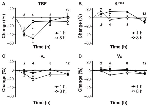
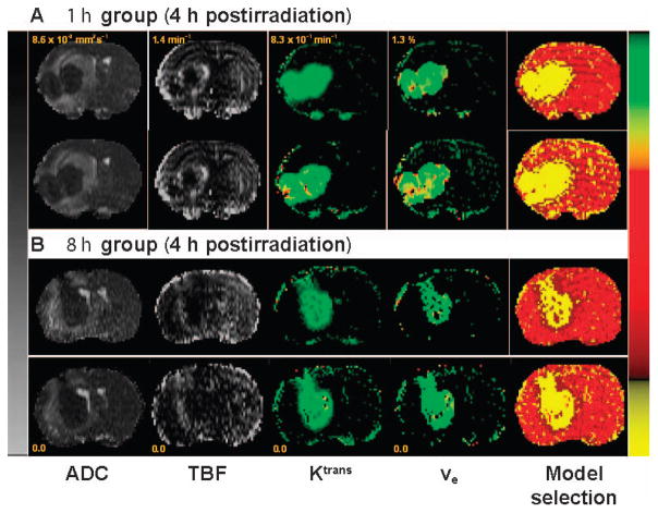
In this study we used magnetic resonance imaging (MRI) biomarkers to monitor the acute temporal changes in tumor vascular physiology with the aim of identifying the vascular signatures that predict response to combined anti-angiogenic and radiation treatments. Forty-three athymic rats implanted with orthotopic U-251 glioma cells were studied for approximately 21 days after implantation. Two MRI studies were performed on each animal, pre- and post-treatment, to measure tumor vascular parameters. Two animal groups received treatment comprised of Cilengitide, an anti-angiogenic agent and radiation. The first group received a subcurative regimen of Cilengitide 1 h before irradiation, while the second group received a curative regimen of Cilengitide 8 h before irradiation. Cilengitide was given as a single dose (4 mg/kg; intraperitoneal) after the pretreatment MRI study and before receiving a 20 Gy radiation dose. After irradiation, the post-treatment MRI study was performed at selected time points: 2, 4, 8 and 12 h (n = ≥5 per time point). Significant changes in vascular parameters were observed at early time points after combined treatments in both treatment groups (1 and 8 h). The temporal changes in vascular parameters in the first group (treated 1 h before exposure) resembled a previously reported pattern associated with radiation exposure alone. Conversely, in the second group (treated 8 h before exposure), all vascular parameters showed an initial response at 2-4 h postirradiation, followed by an apparent lack of response at later time points. The signature time point to define the "synergy" of Cilengitide and radiation was 4 h postirradiation. For example, 4 h after combined treatments using a 1 h separation (which followed the subcurative regimen), tumor blood flow was significantly decreased, nearly 50% below baseline (P = 0.007), whereas 4 h after combined treatments using an 8 h separation (which followed the curative regimen), tumor blood flow was only 10% less than baseline. Comparison between the first and second groups further revealed that most other vascular parameters were maximally different 4 h after combined treatments. In conclusion, the data are consistent with the assertion that the delivery of radiation at the vascular normalization time window of Cilengitide improves radiation treatment outcome. The different vascular responses after the different delivery times of combined treatments in light of the known tumor responses under similar conditions would indicate that timing has a crucial influence on treatment outcome and long-term survival. Tracking acute changes in tumor physiology after monotherapy or combined treatments appears to aid in identifying the beneficial timing for administration, and perhaps has predictive value. Therefore, judicial timing of treatments may result in optimal treatment response.

https://cdn.ncbi.nlm.nih.gov/pmc/blobs/7b27/5381817/02ccdcbff9fb/nihms848332f5.jpg
https://cdn.ncbi.nlm.nih.gov/pmc/blobs/7b27/5381817/f75e46388861/nihms848332f1.jpg
https://cdn.ncbi.nlm.nih.gov/pmc/blobs/7b27/5381817/6b32a9117abc/nihms848332f2.jpg
https://cdn.ncbi.nlm.nih.gov/pmc/blobs/7b27/5381817/c4188b116429/nihms848332f3.jpg
https://cdn.ncbi.nlm.nih.gov/pmc/blobs/7b27/5381817/56251f60f672/nihms848332f4.jpg
https://cdn.ncbi.nlm.nih.gov/pmc/blobs/7b27/5381817/02ccdcbff9fb/nihms848332f5.jpg
摘要

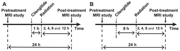
在本研究中,我们使用磁共振成像(MRI)生物标志物来监测肿瘤血管生理学的急性时间变化,目的是识别预测联合抗血管生成和放射治疗反应的血管特征。对43只植入原位U-251胶质瘤细胞的无胸腺大鼠在植入后约21天进行研究。在治疗前和治疗后对每只动物进行两项MRI研究,以测量肿瘤血管参数。两个动物组接受由西仑吉肽(一种抗血管生成药物)和放射组成的治疗。第一组在照射前1小时接受亚治愈剂量的西仑吉肽,而第二组在照射前8小时接受治愈剂量的西仑吉肽。在预处理MRI研究后且在接受20 Gy放射剂量前,西仑吉肽作为单剂量(4 mg/kg;腹腔内注射)给药。照射后,在选定的时间点进行治疗后MRI研究:2、4、8和12小时(每个时间点n≥5)。在两个治疗组的联合治疗后的早期时间点(1和8小时)观察到血管参数有显著变化。第一组(在照射前1小时治疗)的血管参数的时间变化类似于先前报道的仅与放射暴露相关的模式。相反,在第二组(在照射前8小时治疗)中,所有血管参数在照射后2-4小时显示出初始反应,随后在后期时间点明显缺乏反应。定义西仑吉肽和放射“协同作用”的标志性时间点是照射后4小时。例如,在采用1小时间隔的联合治疗后4小时(遵循亚治愈方案),肿瘤血流显著降低,比基线低近50%(P = 0.007),而在采用8小时间隔的联合治疗后4小时(遵循治愈方案),肿瘤血流仅比基线低10%。第一组和第二组之间的比较进一步显示,在联合治疗后4小时,大多数其他血管参数差异最大。总之,数据与以下观点一致,即在西仑吉肽的血管正常化时间窗进行放射治疗可改善放射治疗结果。鉴于在相似条件下已知的肿瘤反应,联合治疗不同给药时间后的不同血管反应表明时间对治疗结果和长期生存有至关重要的影响。追踪单一疗法或联合治疗后肿瘤生理学的急性变化似乎有助于确定有益的给药时间,也许具有预测价值。因此,合理的治疗时间可能导致最佳的治疗反应。

相似文献

1
Acute Temporal Changes of MRI-Tracked Tumor Vascular Parameters after Combined Anti-angiogenic and Radiation Treatments in a Rat Glioma Model: Identifying Signatures of Synergism.大鼠胶质瘤模型中联合抗血管生成与放射治疗后MRI追踪的肿瘤血管参数的急性时间变化:确定协同作用的特征
Radiat Res. 2017 Jan;187(1):79-88. doi: 10.1667/RR14358.1. Epub 2016 Dec 21.
2
Cilengitide-induced temporal variations in transvascular transfer parameters of tumor vasculature in a rat glioma model: identifying potential MRI biomarkers of acute effects.西仑吉肽诱导大鼠胶质瘤模型中肿瘤血管跨血管转运参数的时间变化:确定急性效应的潜在MRI生物标志物
PLoS One. 2013 Dec 23;8(12):e84493. doi: 10.1371/journal.pone.0084493. eCollection 2013.
3
Cilengitide targeting of alpha(v)beta(3) integrin receptor synergizes with radioimmunotherapy to increase efficacy and apoptosis in breast cancer xenografts.西仑吉肽靶向α(v)β(3)整合素受体与放射免疫疗法协同作用,以提高乳腺癌异种移植瘤的疗效并增加细胞凋亡。
Cancer Res. 2002 Aug 1;62(15):4263-72.
4
MRI-Tracked Tumor Vascular Changes in the Hours after Single-Fraction Irradiation.单次分割照射后数小时内MRI追踪的肿瘤血管变化
Radiat Res. 2015 Jun;183(6):713-21. doi: 10.1667/RR13458.1. Epub 2015 May 26.
5
Radiation sensitization of glioblastoma by cilengitide has unanticipated schedule-dependency.西仑吉肽对胶质母细胞瘤的放射增敏作用具有意外的时间依赖性。
Int J Cancer. 2009 Jun 1;124(11):2719-27. doi: 10.1002/ijc.24240.
6
Mechanisms of tumor development and anti-angiogenic therapy in glioblastoma multiforme.多形性胶质母细胞瘤的肿瘤发展机制及抗血管生成治疗
Neurol Med Chir (Tokyo). 2013;53(11):755-63. doi: 10.2176/nmc.ra2013-0200. Epub 2013 Oct 25.
7
A safety run-in and randomized phase 2 study of cilengitide combined with chemoradiation for newly diagnosed glioblastoma (NABTT 0306).一项新诊断胶质母细胞瘤(NABTT 0306)中西仑吉肽联合放化疗的安全性预试验和随机 2 期研究。
Cancer. 2012 Nov 15;118(22):5601-7. doi: 10.1002/cncr.27585. Epub 2012 Apr 19.
8
MRI biomarkers identify the differential response of glioblastoma multiforme to anti-angiogenic therapy.MRI 生物标志物可识别多形性胶质母细胞瘤对抗血管生成治疗的差异化反应。
Neuro Oncol. 2014 Jun;16(6):868-79. doi: 10.1093/neuonc/nou040. Epub 2014 Apr 23.
9
In vitro study of combined cilengitide and radiation treatment in breast cancer cell lines.体外研究联合西利单抗和放射治疗在乳腺癌细胞系中的作用。
Radiat Oncol. 2013 Oct 23;8:246. doi: 10.1186/1748-717X-8-246.
10
Dual inhibition of PFKFB3 and VEGF normalizes tumor vasculature, reduces lactate production, and improves chemotherapy in glioblastoma: insights from protein expression profiling and MRI.双重抑制 PFKFB3 和 VEGF 可使肿瘤血管正常化,减少乳酸生成,并改善胶质母细胞瘤的化疗效果:来自蛋白表达谱和 MRI 的见解。
Theranostics. 2020 Jun 5;10(16):7245-7259. doi: 10.7150/thno.44427. eCollection 2020.

引用本文的文献

1
Radiomics characterization of tissues in an animal brain tumor model imaged using dynamic contrast enhanced (DCE) MRI.利用动态对比增强(DCE)MRI 对动物脑肿瘤模型中的组织进行放射组学特征分析。
Sci Rep. 2023 Jul 2;13(1):10693. doi: 10.1038/s41598-023-37723-8.
2
Dynamic contrast enhanced (DCE) MRI estimation of vascular parameters using knowledge-based adaptive models.基于知识的自适应模型的动态对比增强(DCE)MRI 血管参数估计。
Sci Rep. 2023 Jun 14;13(1):9672. doi: 10.1038/s41598-023-36483-9.
3
Characterization of the Response of 9L and U-251N Orthotopic Brain Tumors to 3D Conformal Radiation Therapy.

本文引用的文献

1
MRI-Tracked Tumor Vascular Changes in the Hours after Single-Fraction Irradiation.单次分割照射后数小时内MRI追踪的肿瘤血管变化
Radiat Res. 2015 Jun;183(6):713-21. doi: 10.1667/RR13458.1. Epub 2015 May 26.
2
Cilengitide combined with standard treatment for patients with newly diagnosed glioblastoma with methylated MGMT promoter (CENTRIC EORTC 26071-22072 study): a multicentre, randomised, open-label, phase 3 trial.西仑吉肽联合标准治疗用于伴有 MGMT 启动子甲基化的新诊断胶质母细胞瘤患者(CENTRIC EORTC 26071-22072 研究):一项多中心、随机、开放标签、3 期临床试验。
Lancet Oncol. 2014 Sep;15(10):1100-8. doi: 10.1016/S1470-2045(14)70379-1. Epub 2014 Aug 19.
3
9L 和 U-251N 原位脑肿瘤对 3D 适形放射治疗的反应特性。
Radiat Res. 2023 Mar 1;199(3):217-228. doi: 10.1667/RADE-22-00048.1.
4
A computational model of glioma reveals opposing, stiffness-sensitive effects of leaky vasculature and tumor growth on tissue mechanical stress and porosity.脑胶质瘤的计算模型揭示了渗漏血管和肿瘤生长对组织力学应力和孔隙率的相反的、对刚性敏感的影响。
Biomech Model Mechanobiol. 2021 Oct;20(5):1981-2000. doi: 10.1007/s10237-021-01488-8. Epub 2021 Aug 7.
5
Imaging acute effects of bevacizumab on tumor vascular kinetics in a preclinical orthotopic model of U251 glioma.在 U251 神经胶质瘤的临床前原位模型中,成像贝伐单抗对肿瘤血管动力学的急性影响。
NMR Biomed. 2021 Jul;34(7):e4516. doi: 10.1002/nbm.4516. Epub 2021 Apr 4.
6
The impact of initial tumor microenvironment on imaging phenotype.初始肿瘤微环境对影像学表现的影响。
Cancer Treat Res Commun. 2021;27:100315. doi: 10.1016/j.ctarc.2021.100315. Epub 2021 Jan 19.
7
Toward a noninvasive estimate of interstitial fluid pressure by dynamic contrast-enhanced MRI in a rat model of cerebral tumor.通过动态对比增强 MRI 对脑肿瘤大鼠模型进行无创性间质液压力估计。
Magn Reson Med. 2018 Nov;80(5):2040-2052. doi: 10.1002/mrm.27163. Epub 2018 Mar 9.
8
Reproducibility and relative stability in magnetic resonance imaging indices of tumor vascular physiology over a period of 24h in a rat 9L gliosarcoma model.在大鼠9L胶质肉瘤模型中,肿瘤血管生理学的磁共振成像指标在24小时内的可重复性和相对稳定性。
Magn Reson Imaging. 2017 Dec;44:131-139. doi: 10.1016/j.mri.2017.09.003. Epub 2017 Sep 5.
Intratumor distribution and test-retest comparisons of physiological parameters quantified by dynamic contrast-enhanced MRI in rat U251 glioma.
大鼠U251胶质瘤中通过动态对比增强磁共振成像定量的生理参数的瘤内分布及重测比较。
NMR Biomed. 2014 Oct;27(10):1230-8. doi: 10.1002/nbm.3178. Epub 2014 Aug 14.
4
Axitinib sensitization of high Single Dose Radiotherapy.阿昔替尼对高单次剂量放疗的增敏作用。
Radiother Oncol. 2014 Apr;111(1):88-93. doi: 10.1016/j.radonc.2014.02.010. Epub 2014 Apr 29.
5
Bevacizumab in glioblastoma--still much to learn.贝伐单抗治疗胶质母细胞瘤——仍有很多需要了解的地方。
N Engl J Med. 2014 Feb 20;370(8):764-5. doi: 10.1056/NEJMe1313309.
6
Bevacizumab plus radiotherapy-temozolomide for newly diagnosed glioblastoma.贝伐珠单抗联合放疗-替莫唑胺治疗新诊断的胶质母细胞瘤。
N Engl J Med. 2014 Feb 20;370(8):709-22. doi: 10.1056/NEJMoa1308345.
7
A randomized trial of bevacizumab for newly diagnosed glioblastoma.贝伐珠单抗治疗新诊断的胶质母细胞瘤的随机试验。
N Engl J Med. 2014 Feb 20;370(8):699-708. doi: 10.1056/NEJMoa1308573.
8
Cilengitide-induced temporal variations in transvascular transfer parameters of tumor vasculature in a rat glioma model: identifying potential MRI biomarkers of acute effects.西仑吉肽诱导大鼠胶质瘤模型中肿瘤血管跨血管转运参数的时间变化:确定急性效应的潜在MRI生物标志物
PLoS One. 2013 Dec 23;8(12):e84493. doi: 10.1371/journal.pone.0084493. eCollection 2013.
9
Combining molecular targeted agents with radiation therapy for malignant gliomas.将分子靶向药物与放射治疗联合用于恶性脑胶质瘤。
Onco Targets Ther. 2013 Aug 9;6:1079-95. doi: 10.2147/OTT.S48224. eCollection 2013.
10
Model selection in measures of vascular parameters using dynamic contrast-enhanced MRI: experimental and clinical applications.使用动态对比增强 MRI 进行血管参数测量中的模型选择:实验和临床应用。
NMR Biomed. 2013 Aug;26(8):1028-41. doi: 10.1002/nbm.2996.