Nagaraja Tavarekere N, Aryal Madhava P, Brown Stephen L, Bagher-Ebadian Hassan, Mikkelsen Tom, Yang James J, Panda Swayamprava, Keenan Kelly A, Cabral Glauber, Ewing James R
Department of Anesthesiology, Henry Ford Hospital, Detroit, Michigan, United States of America.
Department of Neurology, Henry Ford Hospital, Detroit, Michigan, United States of America ; Department of Physics, Oakland University, Rochester, Michigan, United States of America.
PLoS One. 2013 Dec 23;8(12):e84493. doi: 10.1371/journal.pone.0084493. eCollection 2013.
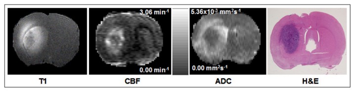
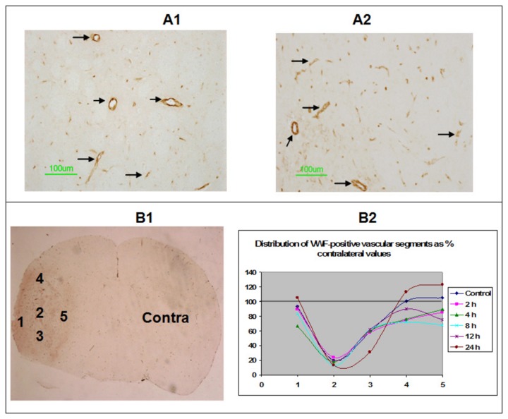
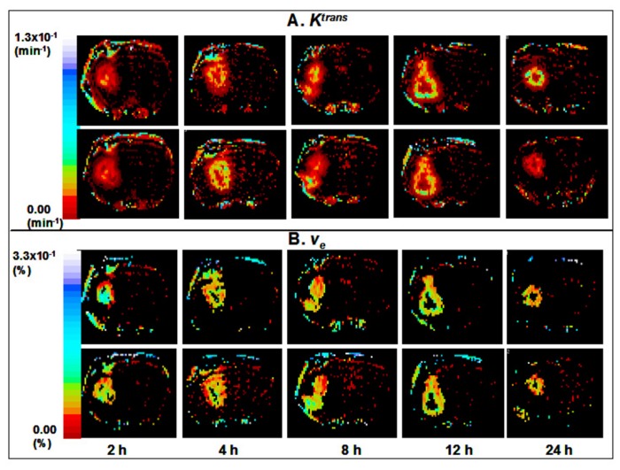
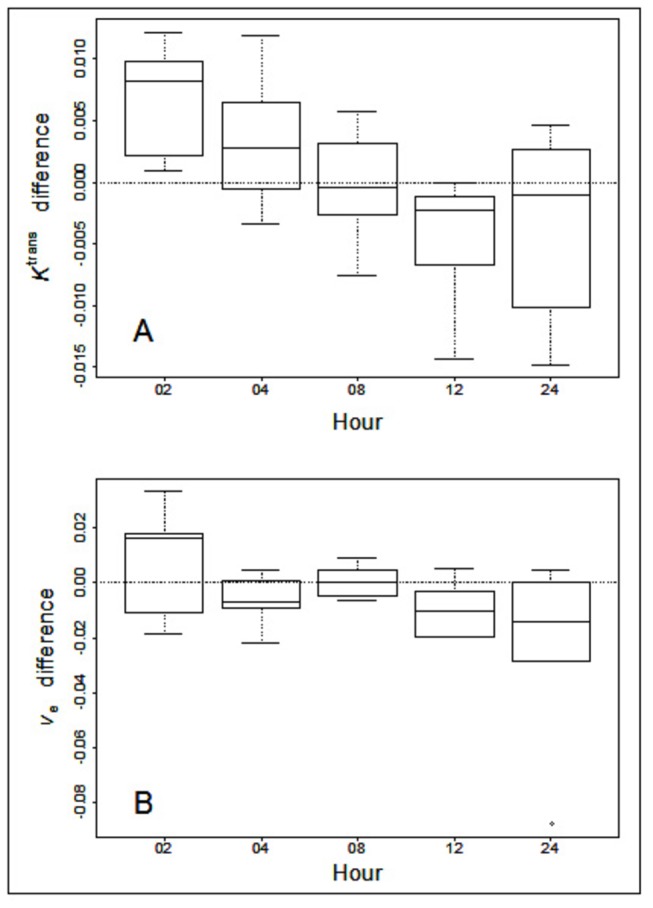
Increased efficacy of radiotherapy (RT) 4-8 h after Cilengitide treatment has been reported. We hypothesized that the effects of Cilengitide on tumor transvascular transfer parameters might underlie, and thus predict, this potentiation. Athymic rats with orthotopic U251 glioma were studied at ~21 days after implantation using dynamic contrast-enhanced (DCE)-MRI. Vascular parameters, viz: plasma volume fraction (v(p)), forward volume transfer constant (K(trans)) and interstitial volume fraction (v(e)) of a contrast agent, were determined in tumor vasculature once before, and again in cohorts 2, 4, 8, 12 and 24 h after Cilengitide administration (4 mg/kg; N = 31; 6-7 per cohort). Perfusion-fixed brain sections were stained for von Willebrand factor to visualize vascular segments. A comparison of pre- and post-treatment parameters showed that the differences between MR indices before and after Cilengitide treatment pivoted around the 8 h time point, with 2 and 4 h groups showing increases, 12 and 24 h groups showing decreases, and values at the 8 h time point close to the baseline. The vascular parameter differences between group of 2 and 4 h and group of 12 and 24 h were significant for K(trans) (p = 0.0001 and v(e) (p = 0,0271). Vascular staining showed little variation with time after Cilengitide. The vascular normalization occurring 8 h after Cilengitide treatment coincided with similar previous reports of increased treatment efficacy when RT followed Cilengitide by 8 h. Pharmacological normalization of vasculature has the potential to increase sensitivity to RT. Evaluating acute temporal responses of tumor vasculature to putative anti-angiogenic drugs may help in optimizing their combination with other treatment modalities.
据报道,西仑吉肽治疗后4 - 8小时放疗(RT)疗效增强。我们推测西仑吉肽对肿瘤跨血管转移参数的影响可能是这种增效作用的基础,从而可以预测这种增效作用。在植入原位U251胶质瘤约21天后,使用动态对比增强(DCE)-MRI对无胸腺大鼠进行研究。在西仑吉肽给药前(4 mg/kg;N = 31;每组6 - 7只)以及给药后2、4、8、12和24小时,分别测定肿瘤血管中的血管参数,即:血浆体积分数(v(p))、正向体积转移常数(K(trans))和造影剂的组织间隙体积分数(v(e))。对灌注固定的脑切片进行血管性血友病因子染色,以观察血管段。治疗前后参数的比较表明,西仑吉肽治疗前后磁共振成像指标的差异在8小时时间点左右出现转折,2小时和4小时组指标升高,12小时和24小时组指标降低,8小时时间点的值接近基线。2小时和4小时组与12小时和24小时组之间的血管参数差异在K(trans)方面具有显著性(p = 0.0001),在v(e)方面也具有显著性(p = 0.0271)。血管染色显示西仑吉肽给药后随时间变化不大。西仑吉肽治疗8小时后出现的血管正常化与之前类似的报道一致,即放疗在西仑吉肽给药8小时后进行时治疗疗效增强。血管的药理学正常化有可能提高对放疗的敏感性。评估肿瘤血管对假定的抗血管生成药物的急性时间反应可能有助于优化它们与其他治疗方式的联合应用。