Märkl Bruno, Wilhelms Narjes, Anthuber Matthias, Schenkirsch Gerhard, Schlimok Günter, Oruzio Daniel
Bruno Märkl, Narjes Wilhelms, Institute of Pathology, Klinikum Augsburg, 86156 Augsburg, Germany.
World J Clin Oncol. 2016 Dec 10;7(6):433-440. doi: 10.5306/wjco.v7.i6.433.
To investigate whether circulating cytokeratin-positive (CK) cells in the mesenteric blood of resected colorectal specimens are prognostic and correlate with tumor budding.
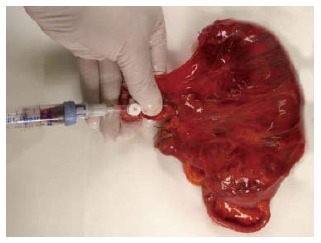
Fifty-six colorectal specimens were collected between 9/2007 and 7/2008. Blood from the mesenteric vein was drawn immediately after receiving the fresh and unfixed specimens in the pathology department. After separation of the mononuclear cells by Ficoll-Hypaque density-gradient centrifugation, cytological smears were immunocytochemically stained for CK18. Tumor budding was evaluated on slides stained for pan-cytokeratin. The identification of ≥ 30 buds/1.3 mm was defined as high grade budding.
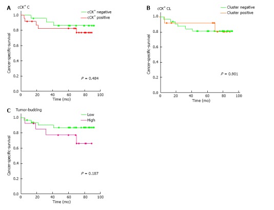
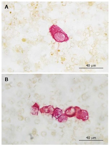
CK cells and clusters were identified in 29 (48%) and 14 (25%) of the samples, respectively. Two cells were identified in one of three non-malignant cases. Clusters were found exclusively in malignant cases. The occurrence of CK cells or clusters was not associated with any of the evaluated clinicopathological factors, including surgical technique and tumor budding. Moreover, the occurrence of CK cells or clusters had no influence on the cancer-specific survival [75 mo (CI: 61; 88) 83 mo (CI: 72; 95) and 80 mo (CI: 63; 98) 79 mo (CI: 69; 89), respectively].
CK cells and showed neither prognostic significance nor an association with tumor budding. It is very likely that CK18-staining is not specific enough to identify the relevant cells.
研究切除的结直肠标本肠系膜血中循环细胞角蛋白阳性(CK)细胞是否具有预后价值以及是否与肿瘤芽生相关。
2007年9月至2008年7月收集了56份结直肠标本。在病理科接收新鲜未固定标本后,立即从肠系膜静脉采血。通过Ficoll-Hypaque密度梯度离心分离单核细胞后,对细胞涂片进行CK18免疫细胞化学染色。在全细胞角蛋白染色的玻片上评估肿瘤芽生情况。将≥30个芽/1.3 mm定义为高级别芽生。
分别在29份(48%)和14份(25%)样本中鉴定出CK细胞和细胞簇。在3例非恶性病例中的1例中发现了2个细胞。细胞簇仅在恶性病例中发现。CK细胞或细胞簇的出现与任何评估的临床病理因素均无关,包括手术技术和肿瘤芽生。此外,CK细胞或细胞簇的出现对癌症特异性生存无影响[分别为75个月(CI:61;88)对83个月(CI:72;95)以及80个月(CI:63;98)对79个月(CI:69;89)]。
CK细胞既无预后意义,也与肿瘤芽生无关。很可能CK18染色不足以特异性地识别相关细胞。