Garcia-Tutor Emilio, Romeo Marco, Chae Michael P, Hunter-Smith David J, Rozen Warren Matthew
Department of Plastic and Reconstructive Surgery, Hospital de Guadalajara, Guadalajara, Spain; MD Anderson Cancer Center, Madrid, Spain.
Department of Plastic and Reconstructive Surgery, Hospital de Guadalajara , Guadalajara , Spain.
Front Surg. 2016 Dec 13;3:66. doi: 10.3389/fsurg.2016.00066. eCollection 2016.
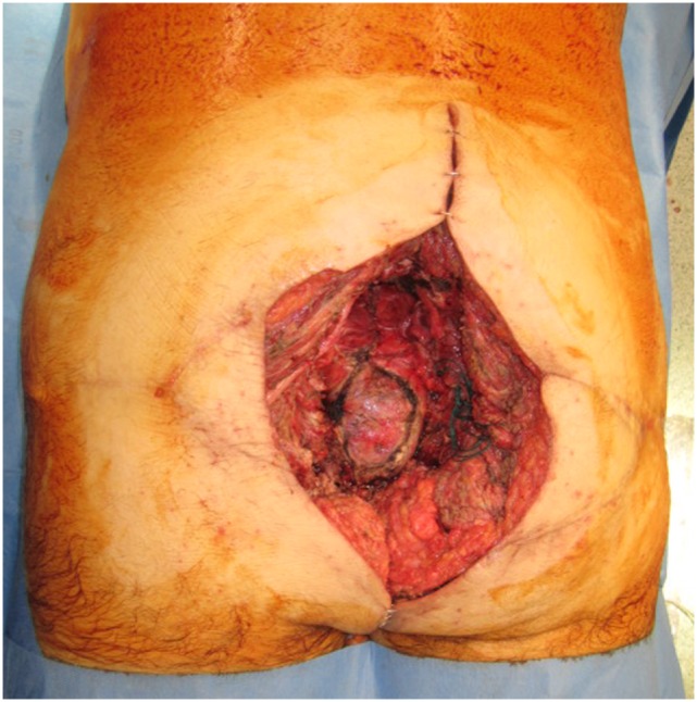
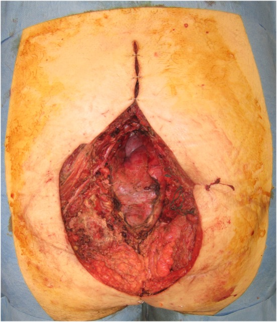
Locoregional flaps are sufficient in most sacral reconstructions. However, large sacral defects due to malignancy necessitate a different reconstructive approach, with local flaps compromised by radiation and regional flaps inadequate for broad surface areas or substantial volume obliteration. In this report, we present our experience using free muscle transfer for volumetric reconstruction, in such cases, and demonstrate three-dimensional (3D) haptic models of the sacral defect to aid preoperative planning.
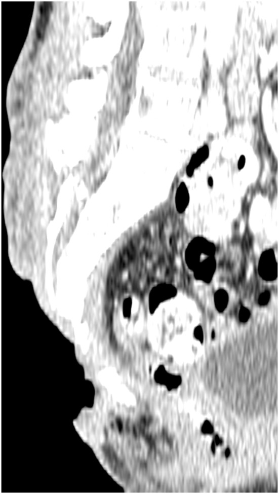
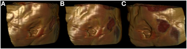
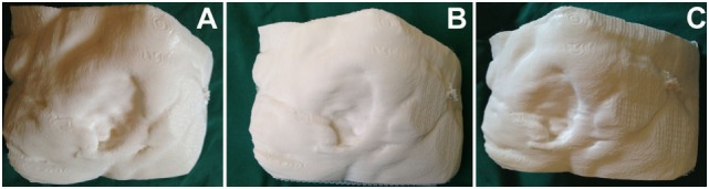
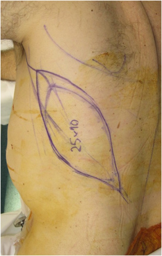
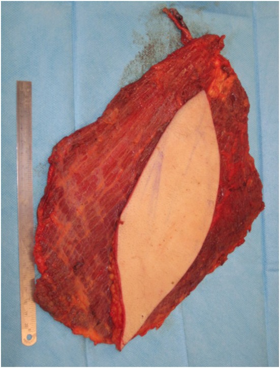
Five consecutive patients with irradiated sacral defects secondary to oncologic resections were included, surface area ranging from 143-600 cm. Latissimus dorsi (LD)-based free flap sacral reconstruction was performed in each case, between 2005 and 2011. Where the superior gluteal artery was compromised, the subcostal artery (SA) was used as a recipient vessel. Microvascular technique, complications, and outcomes are reported. The use of volumetric analysis and 3D printing is also demonstrated, with imaging data converted to 3D images suitable for 3D printing with Osirix software (Pixmeo, Geneva, Switzerland). An office-based, desktop 3D printer was used to print 3D models of sacral defects, used to demonstrate surface area and contour and produce a volumetric print of the dead space needed for flap obliteration.
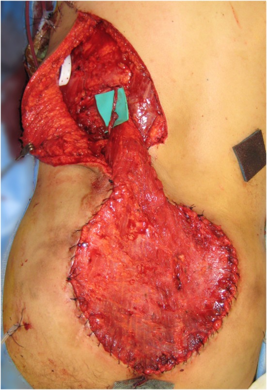
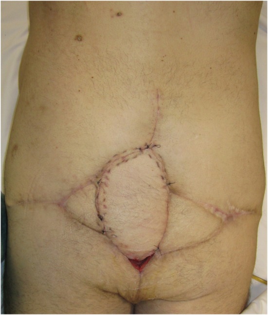
The clinical series of LD free flap reconstructions is presented, with successful transfer in all cases, and adequate soft-tissue cover and volume obliteration achieved. The original use of the SA as a recipient vessel was successfully achieved. All wounds healed uneventfully. 3D printing is also demonstrated as a useful tool for 3D evaluation of volume and dead space.
Free flaps offer unique benefits in sacral reconstruction where local tissue is compromised by irradiation and tumor recurrence, and dead space requires accurate volumetric reconstruction. We describe for the first time the use of the SA as a recipient in free flap sacral reconstruction. 3D printing of haptic bio-models is a rapidly evolving field with a substantial role in preoperative planning.
在大多数骶骨重建手术中,局部区域皮瓣已足够。然而,恶性肿瘤导致的大型骶骨缺损需要不同的重建方法,局部皮瓣会受到放疗影响,而区域皮瓣对于大面积或大量容积填充又不够用。在本报告中,我们介绍了在这类病例中使用游离肌肉移植进行容积重建的经验,并展示了骶骨缺损的三维(3D)触觉模型以辅助术前规划。
纳入连续5例因肿瘤切除导致骶骨缺损且接受过放疗的患者,缺损表面积为143 - 600平方厘米。2005年至2011年期间,对每例患者均进行了基于背阔肌(LD)的游离皮瓣骶骨重建。当上臀动脉受损时,将肋下动脉(SA)用作受区血管。报告了微血管技术、并发症及结果。还展示了容积分析和3D打印的应用,利用Osirix软件(瑞士日内瓦Pixmeo公司)将影像数据转换为适合3D打印的3D图像。使用一台办公用桌面3D打印机打印骶骨缺损的3D模型,用于展示表面积和轮廓,并生成皮瓣填充所需死腔的容积打印件。
展示了LD游离皮瓣重建的临床系列病例,所有病例皮瓣移植均成功,实现了充分的软组织覆盖和容积填充。成功实现了最初将SA用作受区血管的操作。所有伤口均顺利愈合。3D打印也被证明是进行容积和死腔3D评估的有用工具。
在局部组织受放疗和肿瘤复发影响且死腔需要精确容积重建的骶骨重建中,游离皮瓣具有独特优势。我们首次描述了在游离皮瓣骶骨重建中使用SA作为受区血管。触觉生物模型的3D打印是一个快速发展的领域,在术前规划中发挥着重要作用。