• 文献检索
  • 文档翻译
  • 深度研究
  • 学术资讯
  • Suppr Zotero 插件Zotero 插件
  • 邀请有礼
  • 套餐&价格
  • 历史记录
应用&插件
Suppr Zotero 插件Zotero 插件浏览器插件Mac 客户端Windows 客户端微信小程序
定价
高级版会员购买积分包购买API积分包
服务
文献检索文档翻译深度研究API 文档MCP 服务
关于我们
关于 Suppr公司介绍联系我们用户协议隐私条款
关注我们

Suppr 超能文献

核心技术专利:CN118964589B侵权必究
粤ICP备2023148730 号-1Suppr @ 2026

文献检索

告别复杂PubMed语法,用中文像聊天一样搜索,搜遍4000万医学文献。AI智能推荐,让科研检索更轻松。

立即免费搜索

文件翻译

保留排版,准确专业,支持PDF/Word/PPT等文件格式,支持 12+语言互译。

免费翻译文档

深度研究

AI帮你快速写综述,25分钟生成高质量综述,智能提取关键信息,辅助科研写作。

立即免费体验

新辅助治疗后使用3D CUBE与2D T2加权技术对直肠癌进行再分期:一项诊断性研究

Rectal cancer restaging using 3D CUBE vs. 2D T2-weighted technique after neoadjuvant therapy: a diagnostic study.

作者信息

Cao Wuteng, Lian Yanbang, Liu Dechao, Li Fangqian, Zhu Pan, Zhou Zhiyang

机构信息

Department of Radiology, Sixth Affiliated Hospital (Gastrointestinal Hospital) of Sun Yat-sen University, Guangzhou, Guangdong, China.

Department of Radiology, Sixth Affiliated Hospital (Gastrointestinal Hospital) of Sun Yat-sen University, Guangzhou, Guangdong, China

出版信息

Gastroenterol Rep (Oxf). 2017 Aug;5(3):226-231. doi: 10.1093/gastro/gow039. Epub 2016 Dec 26.

DOI:10.1093/gastro/gow039
PMID:28025227
原文链接:https://pmc.ncbi.nlm.nih.gov/articles/PMC5554381/
Abstract

OBJECTIVE

This study aimed to compare the accuracy of rectal cancer restaging after neoadjuvant therapy with 3D CUBE sequence with 2D T2-weighted fast spin-echo (FSE) sequence.

METHODS

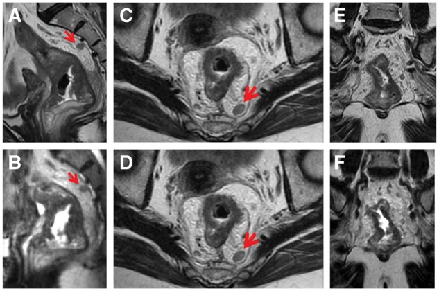
This retrospective study comprised 72 patients with rectal cancer confirmed by colonoscopy and biopsy. After neoadjuvant therapy, all patients underwent pelvic magnetic resonance imaging (MRI) examination at 1.5T MRI sequences including a single coronal 3D CUBE T2-weighted FSE sequence with 1.4 mm thickness and a 2D T2-weighted FSE sequence in the sagittal, coronal and axial planes with 5 mm thickness. The total acquisition time of the two sequences was recorded. Results were compared with postsurgical pathology (gold standard). The diagnostic accuracy was evaluated; and receiver operating characteristic (ROC) curves and the area under the curves (AUC) were calculated.

RESULTS

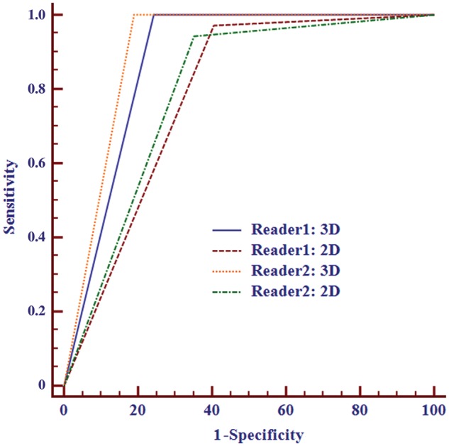
The T category staging accuracy of 3D T2WI and 2D T2WI was 81.9% and 72.2%, respectively, for reviewer 1 and 86.1% and 75.0% for reviewer 2. The AUC of 3D was higher than that of 2D (0.878 vs. 0.783 for reader 1 and 0.905 vs. 0.796 for reader 2; both P < 0.05) when judging whether the tumor broke through the muscle layer. There was no significant difference between 3D and 2D in judging whether lymph nodes were malignant (AUC 0.719 vs. 0.698 for reader 1 and 0.740 vs. 0.698 for reader 2; both P > 0.05). There were no significant differences in the visibility of the rectal wall layer, tumor lesion and the overall image quality (all P > 0.05). Compared with 2D sequences, the 3D sequence had shorter acquisition time and higher signal intensity ratio (both P < 0.05).

CONCLUSION

3D CUBE T2-weighted sequences offer better diagnostic accuracy in rectal cancer restaging after neoadjuvant therapy when compared with 2D T2-weighted FSE sequences; it has a shorter scanning time and more versatility of orientation reconstruction.

摘要

目的

本研究旨在比较三维(3D)CUBE序列与二维(2D)T2加权快速自旋回波(FSE)序列在新辅助治疗后直肠癌再分期中的准确性。

方法

本回顾性研究纳入72例经结肠镜检查及活检确诊的直肠癌患者。新辅助治疗后,所有患者均在1.5T磁共振成像(MRI)序列下行盆腔MRI检查,包括层厚1.4 mm的单次冠状位3D CUBE T2加权FSE序列,以及矢状位、冠状位和轴位上层厚5 mm的2D T2加权FSE序列。记录两种序列的总采集时间。将结果与术后病理(金标准)进行比较。评估诊断准确性;计算受试者工作特征(ROC)曲线及曲线下面积(AUC)。

结果

对于审阅者1,3D T2WI和2D T2WI的T分期准确率分别为81.9%和72.2%,对于审阅者2分别为86.1%和75.0%。在判断肿瘤是否突破肌层时,3D序列的AUC高于2D序列(审阅者1:0.878对0.783;审阅者2:0.905对0.796;P均<0.05)。在判断淋巴结是否为恶性方面,3D与2D之间无显著差异(审阅者1:AUC 0.719对0.698;审阅者2:AUC 0.740对0.698;P均>0.05)。直肠壁层、肿瘤病变的显示及整体图像质量方面均无显著差异(P均>0.05)。与2D序列相比,3D序列采集时间更短,信号强度比更高(P均<0.05)。

结论

与2D T2加权FSE序列相比,3D CUBE T2加权序列在新辅助治疗后直肠癌再分期中具有更好的诊断准确性;扫描时间更短,方向重建更具灵活性。

https://cdn.ncbi.nlm.nih.gov/pmc/blobs/3a19/5554381/be94bbdaaa41/gow039f3.jpg
https://cdn.ncbi.nlm.nih.gov/pmc/blobs/3a19/5554381/81c2728070c3/gow039f1.jpg
https://cdn.ncbi.nlm.nih.gov/pmc/blobs/3a19/5554381/be94bbdaaa41/gow039f3.jpg
https://cdn.ncbi.nlm.nih.gov/pmc/blobs/3a19/5554381/81c2728070c3/gow039f1.jpg
https://cdn.ncbi.nlm.nih.gov/pmc/blobs/3a19/5554381/be94bbdaaa41/gow039f3.jpg

相似文献

1
Rectal cancer restaging using 3D CUBE vs. 2D T2-weighted technique after neoadjuvant therapy: a diagnostic study.新辅助治疗后使用3D CUBE与2D T2加权技术对直肠癌进行再分期:一项诊断性研究
Gastroenterol Rep (Oxf). 2017 Aug;5(3):226-231. doi: 10.1093/gastro/gow039. Epub 2016 Dec 26.
2
Locally advanced rectal cancer: MR imaging for restaging after neoadjuvant radiation therapy with concomitant chemotherapy. Part II. What are the criteria to predict involved lymph nodes?局部进展期直肠癌:新辅助放疗联合化疗后再分期的磁共振成像。第二部分。预测淋巴结受累的标准是什么?
Radiology. 2009 Jul;252(1):81-91. doi: 10.1148/radiol.2521081364. Epub 2009 Apr 29.
3
Three-dimensional isotropic T2-weighted fast spin-echo (VISTA) ankle MRI versus two-dimensional fast spin-echo T2-weighted sequences for the evaluation of anterior talofibular ligament injury.三维各向同性T2加权快速自旋回波(VISTA)踝关节磁共振成像与二维快速自旋回波T2加权序列在评估距腓前韧带损伤中的比较
Clin Radiol. 2016 Apr;71(4):349-55. doi: 10.1016/j.crad.2015.12.007. Epub 2016 Jan 14.
4
Preoperative 3T MR imaging of rectal cancer: local staging accuracy using a two-dimensional and three-dimensional T2-weighted turbo spin echo sequence.直肠癌的术前3T磁共振成像:使用二维和三维T2加权快速自旋回波序列的局部分期准确性
Eur J Radiol. 2008 Jan;65(1):66-71. doi: 10.1016/j.ejrad.2007.11.029. Epub 2007 Dec 27.
5
Image Quality Assessment of 2D 3D T2WI and Evaluation of Ultra-high b-Value (b=2,000 mm/s) DWI for Response Assessment in Rectal Cancer.二维及三维T2WI图像质量评估与超高b值(b = 2000mm²/s)弥散加权成像在直肠癌疗效评估中的应用
Anticancer Res. 2018 Feb;38(2):969-978. doi: 10.21873/anticanres.12311.
6
3D isotropic T2-weighted fast spin echo (VISTA) versus 2D T2-weighted fast spin echo in evaluation of the calcaneofibular ligament in the oblique coronal plane.在斜冠状面评估跟腓韧带时,三维各向同性T2加权快速自旋回波(VISTA)与二维T2加权快速自旋回波的比较
Clin Radiol. 2017 Feb;72(2):176.e1-176.e7. doi: 10.1016/j.crad.2016.09.023. Epub 2016 Nov 9.
7
Comparison of oblique coronal images in knee of three-dimensional isotropic T-weighted turbo spin echo MRI versus two-dimensional fast spin echo T-weighted sequences for evaluation of posterior cruciate ligament injury.三维各向同性T加权快速自旋回波MRI与二维快速自旋回波T加权序列膝关节斜冠状位图像在评估后交叉韧带损伤中的比较。
Br J Radiol. 2016 Nov;89(1067):20160554. doi: 10.1259/bjr.20160554. Epub 2016 Sep 21.
8
MRI of the anterior talofibular ligament, talar cartilage and os subfibulare: Comparison of isotropic resolution 3D and conventional 2D T2-weighted fast spin-echo sequences at 3.0 T.距腓前韧带、距骨软骨和腓骨下骨的磁共振成像:3.0T下各向同性分辨率三维与传统二维T2加权快速自旋回波序列的比较
Skeletal Radiol. 2016 Jul;45(7):899-908. doi: 10.1007/s00256-016-2367-x. Epub 2016 Mar 19.
9
Three-dimensional isotropic T2-weighted fast spin-echo (VISTA) knee MRI at 3.0 T in the evaluation of the anterior cruciate ligament injury with additional views: comparison with two-dimensional fast spin-echo T2-weighted sequences.3.0T三维各向同性T2加权快速自旋回波(VISTA)膝关节MRI在评估前交叉韧带损伤中的应用及附加视图:与二维快速自旋回波T2加权序列的比较
Acta Radiol. 2016 Nov;57(11):1372-1379. doi: 10.1177/0284185114568048. Epub 2016 Jul 21.
10
Rectal cancer: comparison of accuracy of local-regional staging with two- and three-dimensional preoperative 3-T MR imaging.直肠癌:两种和三种术前 3-T MRI 对局部区域分期准确性的比较。
Radiology. 2010 Feb;254(2):485-92. doi: 10.1148/radiol.09090587.

引用本文的文献

1
High-Resolution T2-Weighted MRI to Evaluate Rectal Cancer: Why Variations Matter.高分辨率T2加权磁共振成像评估直肠癌:为何差异至关重要。
Korean J Radiol. 2021 Sep;22(9):1475-1480. doi: 10.3348/kjr.2021.0560.
2
Risk factor analysis for inaccurate pre-operative MRI staging in rectal cancer.直肠癌术前 MRI 分期不准确的危险因素分析。
BMC Cancer. 2020 Mar 27;20(1):253. doi: 10.1186/s12885-020-06761-0.

本文引用的文献

1
MR imaging for rectal cancer: the role in staging the primary and response to neoadjuvant therapy.直肠癌的磁共振成像:在原发性肿瘤分期和新辅助治疗反应中的作用。
Expert Rev Gastroenterol Hepatol. 2014 Aug;8(6):703-19. doi: 10.1586/17474124.2014.906898. Epub 2014 Jun 23.
2
Clinical applications of 3D T2-weighted MRI in pelvic imaging.3D T2加权磁共振成像在盆腔成像中的临床应用。
Abdom Imaging. 2014 Oct;39(5):1052-62. doi: 10.1007/s00261-014-0124-y.
3
Uterine cervical carcinoma: a comparison of two- and three-dimensional T2-weighted turbo spin-echo MR imaging at 3.0 T for image quality and local-regional staging.
子宫颈癌:两种和三种 T2 加权 turbo 自旋回波 MR 成像在 3.0T 下的图像质量和局部区域分期比较。
Eur Radiol. 2013 Apr;23(4):1150-7. doi: 10.1007/s00330-012-2603-2. Epub 2012 Aug 7.
4
Preoperative versus postoperative chemoradiotherapy for locally advanced rectal cancer: results of the German CAO/ARO/AIO-94 randomized phase III trial after a median follow-up of 11 years.局部进展期直肠癌术前与术后放化疗的比较:中位随访 11 年后德国 CAO/ARO/AIO-94 随机 III 期临床试验结果。
J Clin Oncol. 2012 Jun 1;30(16):1926-33. doi: 10.1200/JCO.2011.40.1836. Epub 2012 Apr 23.
5
MR imaging evaluation of perianal fistulas: spectrum of imaging features.磁共振成像评估肛周瘘管:影像学特征谱。
Radiographics. 2012 Jan-Feb;32(1):175-94. doi: 10.1148/rg.321115040.
6
Uterine tumors: comparison of 3D versus 2D T2-weighted turbo spin-echo MR imaging at 3.0 T--initial experience.子宫肿瘤:3.0T 场强下 3D 与 2D T2 加权快速自旋回波 MR 成像的对比——初步经验。
Radiology. 2011 Jan;258(1):154-63. doi: 10.1148/radiol.10100866. Epub 2010 Nov 2.
7
Apparent diffusion coefficient for evaluating tumour response to neoadjuvant chemoradiation therapy for locally advanced rectal cancer.评估局部进展期直肠癌新辅助放化疗疗效的表观扩散系数。
Eur Radiol. 2011 May;21(5):987-95. doi: 10.1007/s00330-010-1989-y. Epub 2010 Oct 27.
8
MRI of the pelvis in women: 3D versus 2D T2-weighted technique.盆腔 MRI:3D 与 2D T2 加权技术比较。
AJR Am J Roentgenol. 2010 Jul;195(1):254-9. doi: 10.2214/ajr.09.3226.
9
Prostate cancer: Comparison of 3D T2-weighted with conventional 2D T2-weighted imaging for image quality and tumor detection.前列腺癌:3D T2 加权成像与传统 2D T2 加权成像在图像质量和肿瘤检测方面的比较。
AJR Am J Roentgenol. 2010 Feb;194(2):446-52. doi: 10.2214/AJR.09.3217.
10
Rectal cancer: comparison of accuracy of local-regional staging with two- and three-dimensional preoperative 3-T MR imaging.直肠癌:两种和三种术前 3-T MRI 对局部区域分期准确性的比较。
Radiology. 2010 Feb;254(2):485-92. doi: 10.1148/radiol.09090587.