Mesarwi Omar A, Shin Mi-Kyung, Bevans-Fonti Shannon, Schlesinger Christina, Shaw Janet, Polotsky Vsevolod Y
Department of Medicine, University of California San Diego School of Medicine, San Diego, California, United States of America.
Department of Medicine, Johns Hopkins University School of Medicine, Baltimore, Maryland, United States of America.
PLoS One. 2016 Dec 28;11(12):e0168572. doi: 10.1371/journal.pone.0168572. eCollection 2016.
Obstructive sleep apnea (OSA) is associated with the progression of non-alcoholic fatty liver disease (NAFLD) to steatohepatitis and fibrosis. This progression correlates with the severity of OSA-associated hypoxia. In mice with diet induced obesity, hepatic steatosis leads to liver tissue hypoxia, which worsens with exposure to intermittent hypoxia. Emerging data has implicated hepatocyte cell signaling as an important factor in hepatic fibrogenesis. We hypothesized that hepatocyte specific knockout of the oxygen sensing α subunit of hypoxia inducible factor-1 (HIF-1), a master regulator of the global response to hypoxia, may be protective against the development of liver fibrosis.
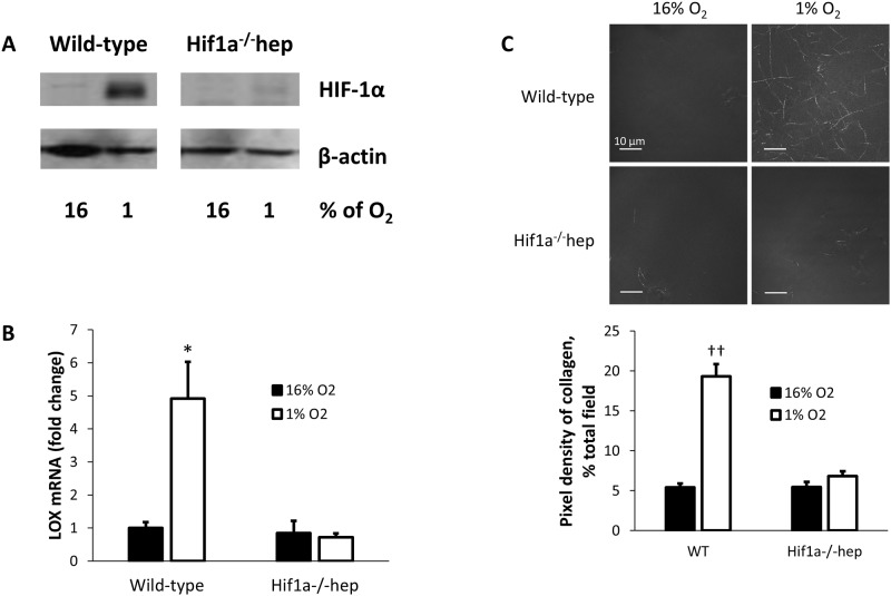
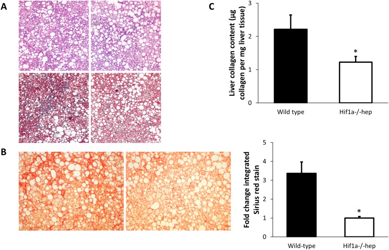
Wild-type mice and mice with hepatocyte-specific HIF-1α knockout (Hif1a-/-hep) were fed a high trans-fat diet for six months, as a model of NAFLD. Hepatic fibrosis was evaluated by Sirius red stain and hydroxyproline assay. Liver enzymes, fasting insulin, and hepatic triglyceride content were also assessed. Hepatocytes were isolated from Hif1a-/-hep mice and wild-type controls and were exposed to sustained hypoxia (1% O2) or normoxia (16% O2) for 24 hours. The culture media was used to reconstitute type I collagen and the resulting matrices were examined for collagen cross-linking.
Wild-type mice on a high trans-fat diet had 80% more hepatic collagen than Hif1a-/-hep mice (2.21 μg collagen/mg liver tissue, versus 1.23 μg collagen/mg liver tissue, p = 0.03), which was confirmed by Sirius red staining. Body weight, liver weight, mean hepatic triglyceride content, and fasting insulin were similar between groups. Culture media from wild-type mouse hepatocytes exposed to hypoxia allowed for avid collagen cross-linking, but very little cross-linking was seen when hepatocytes were exposed to normoxia, or when hepatocytes from Hif1a-/-hep mice were used in hypoxia or normoxia.
Hepatocyte HIF-1 mediates an increase in liver fibrosis in a mouse model of NAFLD, perhaps due to liver tissue hypoxia in hepatic steatosis. HIF-1 is necessary for collagen cross-linking in an in vitro model of fibrosis.
阻塞性睡眠呼吸暂停(OSA)与非酒精性脂肪性肝病(NAFLD)进展为脂肪性肝炎和肝纤维化有关。这种进展与OSA相关缺氧的严重程度相关。在饮食诱导肥胖的小鼠中,肝脏脂肪变性会导致肝组织缺氧,而暴露于间歇性缺氧时情况会恶化。新出现的数据表明,肝细胞信号传导是肝纤维化形成的一个重要因素。我们假设,肝细胞特异性敲除缺氧诱导因子-1(HIF-1)的氧感应α亚基(HIF-1是全局缺氧反应的主要调节因子)可能对肝纤维化的发展具有保护作用。
将野生型小鼠和肝细胞特异性HIF-1α敲除(Hif1a-/-hep)小鼠作为NAFLD模型,喂食高反式脂肪饮食六个月。通过天狼星红染色和羟脯氨酸测定评估肝纤维化。还评估了肝酶、空腹胰岛素和肝脏甘油三酯含量。从Hif1a-/-hep小鼠和野生型对照中分离肝细胞,并将其暴露于持续缺氧(1%氧气)或常氧(16%氧气)24小时。使用培养基重构I型胶原,并检查所得基质的胶原交联情况。
喂食高反式脂肪饮食的野生型小鼠肝脏胶原比Hif1a-/-hep小鼠多80%(2.21μg胶原/毫克肝组织,而Hif1a-/-hep小鼠为1.23μg胶原/毫克肝组织,p = 0.03),天狼星红染色证实了这一点。两组之间的体重、肝脏重量、平均肝脏甘油三酯含量和空腹胰岛素相似。暴露于缺氧的野生型小鼠肝细胞的培养基允许大量胶原交联,但当肝细胞暴露于常氧时,或者当使用Hif1a-/-hep小鼠的肝细胞在缺氧或常氧条件下时,几乎看不到交联。
在NAFLD小鼠模型中,肝细胞HIF-1介导肝纤维化增加,这可能是由于肝脂肪变性中的肝组织缺氧所致。在体外纤维化模型中,HIF-1是胶原交联所必需的。