Kase Satoru, Saito Wataru, Mori Shohei, Saito Michiyuki, Ando Ryo, Dong Zhenyu, Suzuki Tomohiro, Noda Kousuke, Ishida Susumu
Department of Ophthalmology, Hokkaido University Graduate School of Medicine, Sapporo, Japan.
Clin Ophthalmol. 2016 Dec 16;11:9-14. doi: 10.2147/OPTH.S119762. eCollection 2017.
The aims of this study were to analyze optical coherence tomography (OCT) imaging of large macular holes (MHs) treated with inverted internal limiting membrane (ILM) flap technique and to perform a histological examination of an ILM-like membrane tissue obtained during vitrectomy.
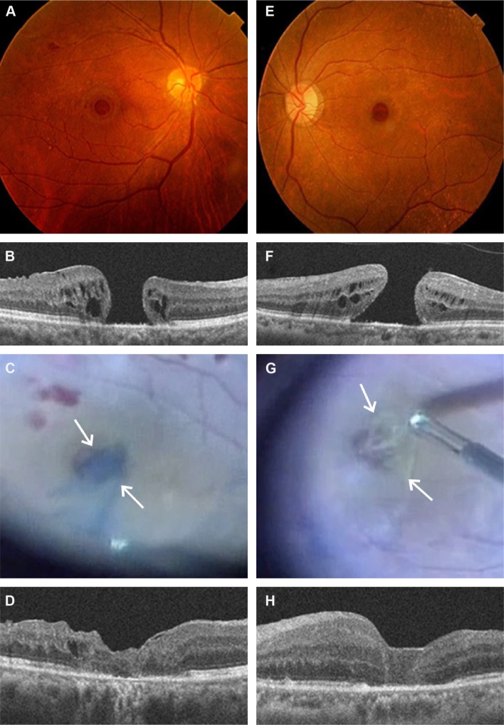
This is a retrospective observational case study. Nine patients, comprising of five males and four females, showing large and myopic MHs, underwent pars plana vitrectomy (PPV) with inverted ILM flap technique assisted by brilliant blue G (BBG) staining. Ophthalmological findings including visual acuity and OCT were investigated based on medical records. Formalin-fixed paraffin-embedded tissue section of an ILM-like membrane was submitted for immunohistochemistry with glial fibrillary acidic protein (GFAP).
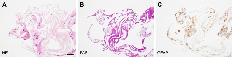
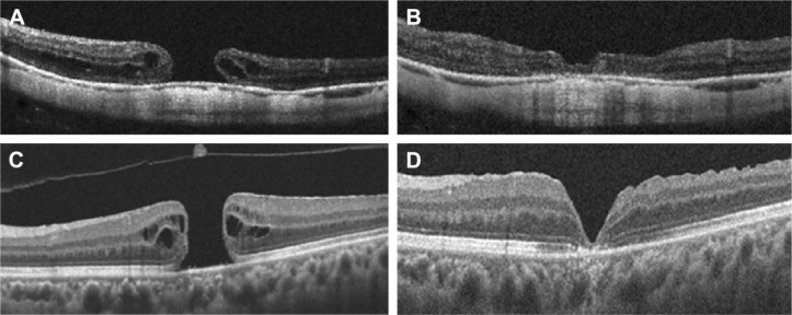
ILM was clearly stained with BBG in eight patients, whereas the ILM in one case revealed no staining with BBG during PPV. Visual acuities improved to >0.2 LogMAR in six patients. The complete closure of MH following PPV with inverted ILM technique was eventually achieved in all patients determined by OCT imaging (100%). Only one patient showed recovery of ellipsoid zone and interdigitation zone following the surgery. Elongation of outer nuclear layer was noted in three eyes. The ILM-like membrane not stained with BBG histologically revealed an amorphous structure admixed with GFAP-positive mononuclear cell infiltration.
PPV with inverted ILM flap technique achieved 100% closure rates with favorable configuration at an initial surgery in large MHs. Our histopathological data also suggest that even BBG staining-negative membrane may be a useful material for autologous transplantation to the hole.
本研究旨在分析采用倒置内界膜(ILM)瓣技术治疗的大黄斑裂孔(MH)的光学相干断层扫描(OCT)成像,并对玻璃体切除术中获得的类ILM膜组织进行组织学检查。
这是一项回顾性观察性病例研究。9例患者,包括5例男性和4例女性,表现为大的近视性MH,接受了在亮蓝G(BBG)染色辅助下的经平坦部玻璃体切除术(PPV)及倒置ILM瓣技术。基于病历调查包括视力和OCT在内的眼科检查结果。将类ILM膜的福尔马林固定石蜡包埋组织切片送去进行胶质纤维酸性蛋白(GFAP)免疫组织化学检测。
8例患者的ILM被BBG清晰染色,而1例患者在PPV期间ILM未被BBG染色。6例患者的视力提高到>0.2 LogMAR。通过OCT成像确定,所有患者最终均通过倒置ILM技术PPV实现了MH的完全闭合(100%)。术后仅1例患者的椭圆体带和指状交叉带恢复。3只眼观察到外核层延长。组织学上未被BBG染色的类ILM膜显示为无定形结构,伴有GFAP阳性单核细胞浸润。
对于大的MH,采用倒置ILM瓣技术的PPV在初次手术时实现了100%的闭合率,且形态良好。我们的组织病理学数据还表明,即使是BBG染色阴性的膜也可能是用于黄斑裂孔自体移植的有用材料。